
See the relationships.
Explore real atlas surfaces: skull and individual sides, muscles of mastication, dental nerves, salivary ducts, vessels, lymph nodes and TMJ structures.
Head and neck anatomy ↗Nerve injury ↗Salivary gland disease ↗
Turn it around. Peel back a layer. Pick a structure and connect what you see to what you know.
Choose a view to set up the model, then try the recall question.
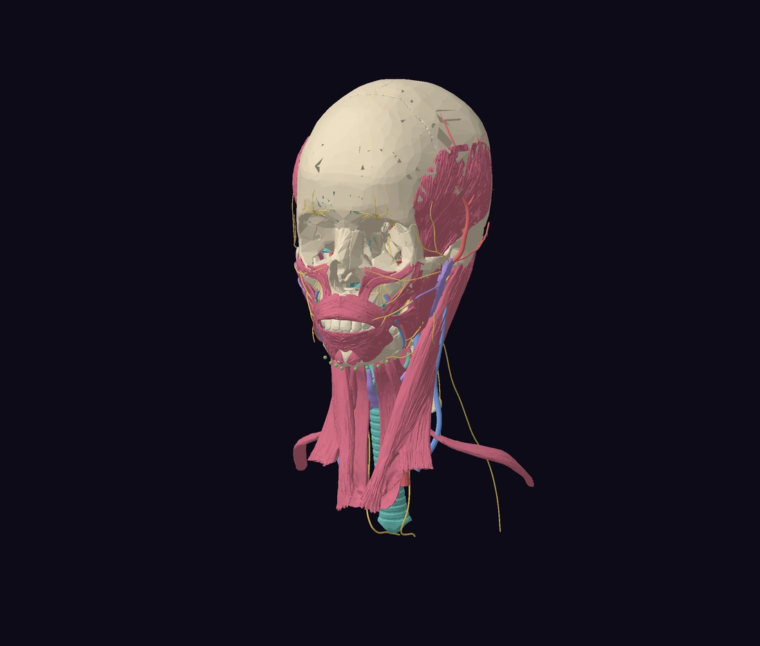
Rotate, zoom and click the model to explore.
The 3D model downloads when you start.
Drag to rotate · scroll or pinch to zoom · right-drag to pan. Keyboard: focus the model, use arrow keys to rotate, +/− to zoom and Home to reset. Choose any structure from the list for the same teaching notes.
Moves a display cut through the outer tooth and ligament. The source pulp cavity stays intact. Cut surfaces are open, without invented tissue layers.
Choose a view, then load the interactive model. The text reference works without 3D.
Detailed Z-Anatomy reference surfaces replace the earlier schematic head overlays. Left and right structures are separately selectable. Hide superficial tissues to expose deeper relationships. The lower neck is regionally cropped; small branches and anatomical variation remain incomplete.
Explore real atlas surfaces: skull and individual sides, muscles of mastication, dental nerves, salivary ducts, vessels, lymph nodes and TMJ structures.
Head and neck anatomy ↗Nerve injury ↗Salivary gland disease ↗A full-body skeleton with selectable organ groups. Relate the heart, lungs, liver and kidneys to dental care.
Immunology and inflammation ↗Acute dental pain ↗Four guided views connect crown and root, hard tissues, pulp space and supporting tissues with short recall questions. Use the tissue guide and a published molar example together.
Dental anatomy and occlusion ↗Oral histology ↗Pulpal and apical diagnosis ↗Periodontal health and gingivitis ↗Select individual adult teeth, check FDI numbering and explore the mandible, maxilla and TMJ relationships.
Dental anatomy and occlusion ↗Occlusion for prosthodontics ↗Simple extraction ↗Use this reference with or without 3D. Names refer to the patient’s right and left. Left and right head structures can be selected separately; some small bones and whole-body structures remain grouped.
Detailed Z-Anatomy reference surfaces replace the earlier schematic head overlays. Left and right structures are separately selectable. Hide superficial tissues to expose deeper relationships. The lower neck is regionally cropped; small branches and anatomical variation remain incomplete.
The lower jaw consists of a body and two rami. Its condylar processes articulate with the temporal bones; the coronoid processes receive temporalis.
Dental connection. Relate the mandibular canal, mental foramen and root positions to assessment before surgery. A surface model cannot establish a patient’s nerve position.
Recall. Which nerve leaves the mental foramen?
Answer. The mental nerve, a terminal branch of the inferior alveolar nerve.
The paired maxillae support the upper teeth and contribute to the hard palate, nasal cavity and orbital floor.
Dental connection. Posterior upper roots may lie close to the maxillary sinus. Their relationship must be assessed on appropriate patient imaging.
Recall. Which bones form most of the anterior hard palate?
Answer. The palatine processes of the maxillae.
The frontal bone forms the forehead, roofs of the orbits and part of the anterior cranial fossa.
Dental connection. The orbital roof helps orientate facial trauma and skull-base anatomy.
Recall. Which cranial fossa lies immediately above the orbital roofs?
Answer. The anterior cranial fossa.
The paired parietal bones form much of the roof and lateral walls of the cranial vault.
Dental connection. Use the cranial vault to orientate superior and lateral skull views.
Recall. Which suture lies between the two parietal bones?
Answer. The sagittal suture.
The temporal bones contain the middle and inner ear regions. Each has a mandibular fossa and articular eminence at the TMJ.
Dental connection. The mandibular condyle articulates with the temporal component through an intervening articular disc.
Recall. Which cranial nerve exits at the stylomastoid foramen?
Answer. The facial nerve, CN VII.
Head and neck anatomy ↗Facial pain and TMD ↗Occlusion for prosthodontics ↗
The occipital bone forms the posterior and inferior cranium. The foramen magnum connects the cranial cavity with the vertebral canal.
Dental connection. The occipital condyles articulate with the atlas.
Recall. Which vertebra articulates directly with the occipital condyles?
Answer. The atlas, C1.
The sphenoid lies centrally in the skull base. Its greater wings, pterygoid processes and foramina connect several important anatomical regions.
Dental connection. Distinguish foramen rotundum for V2 from foramen ovale for V3.
Recall. Which trigeminal division passes through foramen ovale?
Answer. The mandibular division, V3.
The ethmoid contributes to the medial orbital walls, nasal septum and roof of the nasal cavity.
Dental connection. Its cribriform plate transmits olfactory nerve filaments.
Recall. Which special sense is carried by CN I?
Answer. Smell.
The zygomatic bones form the cheek prominences and contribute to the lateral orbital walls and floors.
Dental connection. The zygomatic arch provides attachment for masseter.
Recall. Which muscle arises from the zygomatic arch?
Answer. Masseter.
The horizontal plates of the palatine bones form the posterior part of the hard palate.
Dental connection. Palatal neurovascular bundles travel close to the bony palate. This surface view does not show every foramen.
Recall. Which bones form the posterior hard palate?
Answer. The horizontal plates of the palatine bones.
The paired nasal bones form the bony bridge of the nose.
Dental connection. Their position provides a midline landmark when orientating facial structures.
Recall. Is the entire nasal septum formed by the nasal bones?
Answer. No. The septum includes the vomer, perpendicular plate of the ethmoid and septal cartilage.
The vomer is a thin midline bone forming part of the nasal septum.
Dental connection. Compare it with the ethmoid and the hard palate from an inferior view.
Recall. Which bone forms the posteroinferior bony nasal septum?
Answer. The vomer.
The hyoid is suspended in the upper neck by muscles and ligaments. It provides attachments for tongue, floor-of-mouth and laryngeal muscles.
Dental connection. Use it to understand suprahyoid and infrahyoid muscle groups.
Recall. Does the hyoid articulate directly with another bone?
Answer. No. It is suspended by soft-tissue attachments.
C1 is a ring-shaped vertebra that supports the skull and articulates with the axis.
Dental connection. Its superior facets articulate with the occipital condyles.
Recall. Which movement predominates at the atlanto-occipital joints?
Answer. Flexion and extension, as in nodding.
The dens of C2 acts as a pivot around which the atlas and head rotate.
Dental connection. Distinguish the dens from the body of an ordinary cervical vertebra.
Recall. Which vertebral feature forms the pivot for much of upper cervical rotation?
Answer. The dens of the axis.
The remaining cervical vertebrae support the neck and protect the cervical spinal cord.
Dental connection. Comfortable, supported head and neck positioning matters during dental treatment.
Recall. How many cervical vertebrae are normally present?
Answer. Seven, C1 to C7.
This paired muscle runs from the sternum and clavicle to the mastoid process. Unilateral contraction turns the face towards the opposite side.
Dental connection. It is a key surface landmark separating the anterior and posterior triangles of the neck.
Recall. Which nerve provides its main motor supply?
Answer. The spinal accessory nerve, CN XI.
Each digastric has anterior and posterior bellies connected by an intermediate tendon near the hyoid.
Dental connection. The anterior belly is supplied through V3; the posterior belly is supplied by CN VII.
Recall. Do both digastric bellies have the same motor nerve supply?
Answer. No. The anterior belly receives the nerve to mylohyoid from V3; the posterior belly receives the facial nerve.
The mylohyoids form much of the muscular floor of the mouth between the mandible and hyoid.
Dental connection. The relationship between a root apex and the mylohyoid attachment helps explain routes of odontogenic infection spread.
Recall. Which nerve supplies mylohyoid?
Answer. The nerve to mylohyoid, a branch of the inferior alveolar nerve before it enters the mandibular foramen.
Geniohyoid lies above mylohyoid and runs from the inferior mental spines to the hyoid.
Dental connection. It helps pull the hyoid forwards and upwards during swallowing.
Recall. Which fibres supply geniohyoid?
Answer. C1 fibres travelling with the hypoglossal nerve.
Stylohyoid runs from the styloid process towards the hyoid. It elevates and retracts the hyoid.
Dental connection. Compare its position with the posterior belly of digastric.
Recall. Which cranial nerve supplies stylohyoid?
Answer. The facial nerve, CN VII.
Genioglossus is a fan-shaped extrinsic tongue muscle arising from the superior mental spines.
Dental connection. It contributes to tongue protrusion and helps maintain the tongue’s anterior position.
Recall. Towards which side does the protruded tongue deviate in a unilateral lower motor neurone hypoglossal lesion?
Answer. Towards the affected side.
Hyoglossus runs from the hyoid to the side of the tongue and helps depress the tongue.
Dental connection. The lingual artery passes deep to hyoglossus; the hypoglossal nerve lies superficial to it.
Recall. Is the lingual artery superficial or deep to hyoglossus?
Answer. Deep to hyoglossus.
Sternohyoid runs from the sternum towards the hyoid and helps depress the hyoid.
Dental connection. It belongs to the infrahyoid or strap-muscle group.
Recall. Is sternohyoid a suprahyoid or infrahyoid muscle?
Answer. Infrahyoid.
Omohyoid has two bellies connected by an intermediate tendon, extending between the scapula and hyoid.
Dental connection. Its superior belly forms a boundary within the anterior triangle of the neck.
Recall. How many bellies does omohyoid have?
Answer. Two: superior and inferior.
Sternothyroid extends from the sternum to the thyroid cartilage and helps lower the larynx.
Dental connection. The thyroid cartilage and thyroid gland are different structures.
Recall. Does sternothyroid insert into the hyoid?
Answer. No. It inserts into the thyroid cartilage.
Thyrohyoid connects the thyroid cartilage with the hyoid. It can depress the hyoid or elevate the larynx.
Dental connection. Unlike most strap muscles, its motor supply is C1 carried with CN XII.
Recall. Which cervical root supplies thyrohyoid?
Answer. C1, travelling with the hypoglossal nerve.
The tongue supports speech, swallowing and manipulation of food. Intrinsic muscles change its shape and extrinsic muscles change its position.
Dental connection. Separate general sensation, taste and motor supply when examining tongue symptoms.
Recall. Which nerve carries general sensation from the anterior two-thirds?
Answer. The lingual nerve, from V3. Taste from that region travels in chorda tympani, from CN VII.
The superior, middle and inferior pharyngeal constrictors act during swallowing.
Dental connection. The pharynx is behind the oral and nasal cavities and continues towards the oesophagus.
Recall. What is the principal function of the pharyngeal constrictors?
Answer. To help propel a swallowed bolus towards the oesophagus.
The thyroid cartilage forms a large part of the laryngeal framework, above the cricoid cartilage.
Dental connection. It is part of the airway framework, not the endocrine thyroid gland.
Recall. Which cartilage lies immediately below the thyroid cartilage?
Answer. The cricoid cartilage.
The cricoid is a complete cartilaginous ring below the thyroid cartilage and above the trachea.
Dental connection. Use it to orientate the transition from larynx to trachea.
Recall. Which laryngeal cartilage forms a complete ring?
Answer. The cricoid cartilage.
The epiglottis is an elastic cartilage at the laryngeal inlet. Airway protection during swallowing involves several coordinated actions.
Dental connection. Do not think of the epiglottis as the only mechanism preventing aspiration.
Recall. Where does the epiglottis lie relative to the laryngeal inlet?
Answer. At its anterior margin, extending upwards behind the tongue.
The submandibular glands wrap around the posterior border of mylohyoid, with superficial and deep portions.
Dental connection. Its duct is now separately selectable. Follow the gland around the posterior border of mylohyoid and compare the duct with the floor of the mouth.
Recall. Where does the submandibular duct open?
Answer. At the sublingual caruncle beside the lingual frenulum.
The sublingual glands lie in the anterior floor of the mouth, above mylohyoid and below the mucosa.
Dental connection. A ranula is associated with mucus escape from the sublingual gland, although swelling requires clinical assessment.
Recall. Which major salivary gland sits in the anterior floor of the mouth?
Answer. The sublingual gland.
Each common carotid artery ascends in the neck before dividing into internal and external carotid arteries.
Dental connection. This dataset shows selected vessel segments; a visible gap is not a real anatomical interruption.
Recall. Which carotid branch normally gives no branches in the neck?
Answer. The internal carotid artery.
The internal carotid arteries pass towards the skull base and supply the brain and orbit.
Dental connection. They do not normally supply the superficial face through cervical branches.
Recall. Which carotid artery supplies most external face and jaw structures?
Answer. The external carotid artery, through its branches.
The internal jugular veins are major venous drainage channels from the brain and deep head and neck.
Dental connection. They descend in the carotid sheath alongside the carotid artery and vagus nerve.
Recall. Which cranial nerve travels within the carotid sheath?
Answer. The vagus nerve, CN X.
The upper arch contains incisors, canines, premolars and molars. This reference model includes 14 upper teeth, without third molars.
Dental connection. Switch to Jaws & dentition to select each tooth individually and check its FDI number.
Recall. Which FDI quadrants contain the permanent upper teeth?
Answer. Quadrant 1 is upper right; quadrant 2 is upper left.
The lower permanent teeth are supported by the mandibular alveolar process. This reference model includes 14 lower teeth, without third molars.
Dental connection. Switch to Jaws & dentition to explore each individual tooth and its roots.
Recall. Which FDI quadrants contain the permanent lower teeth?
Answer. Quadrant 3 is lower left; quadrant 4 is lower right.
Masseter runs from the zygomatic arch to the lateral ramus and angle of the mandible. It elevates the jaw.
Dental connection. Motor supply is from V3. This original overlay illustrates the attachment region, not individual muscle fibres.
Recall. Which trigeminal division supplies masseter?
Answer. V3, through the masseteric nerve.
Temporalis has a broad origin in the temporal fossa and a tendon inserting into the coronoid process.
Dental connection. It elevates the mandible; its posterior fibres assist retraction.
Recall. Which mandibular process receives temporalis?
Answer. The coronoid process.
The medial pterygoid attaches to the medial ramus and angle, forming a muscular sling with masseter.
Dental connection. It assists elevation and protrusion. The displayed volume is a schematic attachment-to-attachment guide.
Recall. Which muscle forms the medial part of the pterygomasseteric sling?
Answer. Medial pterygoid.
Lateral pterygoid extends towards the condylar neck and the TMJ disc-capsule complex.
Dental connection. Its actions contribute to protrusion and coordinated opening. This simplified overlay does not separate both heads.
Recall. Which muscle has attachments to the condylar neck and disc-capsule complex?
Answer. Lateral pterygoid.
The parotid glands occupy the region around the posterior ramus and upper masseter, below and in front of each ear.
Dental connection. CN VII passes through the gland, but its secretomotor supply is derived from CN IX via the otic ganglion.
Recall. Where does the parotid duct open into the mouth?
Answer. Opposite the upper second molar.
V2 is the sensory maxillary division of the trigeminal nerve. It leaves the cranial cavity through foramen rotundum.
Dental connection. Its branches supply the upper teeth, palate and midface. This source mesh represents V2; it does not resolve every terminal dental branch.
Recall. Is V2 sensory, motor or mixed?
Answer. Sensory.
V3 is the mixed mandibular division of the trigeminal nerve and leaves the skull through foramen ovale.
Dental connection. It supplies lower facial sensation and motor fibres to the muscles of mastication.
Recall. Which major trigeminal division carries motor fibres?
Answer. V3.
The inferior alveolar nerve is a branch of V3. It enters the mandibular foramen and travels within the mandibular canal.
Dental connection. Trace the source nerve into the mandibular canal, then compare it with the mental branch. Individual root-to-canal relationships require patient imaging.
Recall. Which terminal branch emerges at the mental foramen?
Answer. The mental nerve.
The lingual nerve passes into the floor of the mouth and carries general sensation from the anterior two-thirds of the tongue.
Dental connection. Chorda tympani joins it, carrying taste and preganglionic parasympathetic fibres. Its relation to the lower third molar region varies.
Recall. Which cranial nerve contributes the chorda tympani fibres?
Answer. The facial nerve, CN VII.
The mental nerve leaves the mandible through the mental foramen and supplies the lower lip and chin.
Dental connection. Lower lip or chin sensory change should be recorded carefully and interpreted in its clinical context.
Recall. Does the mental nerve supply tongue sensation?
Answer. No. General sensation to the anterior tongue is carried by the lingual nerve.
The extracranial facial nerve enters the parotid region and divides into branches to muscles of facial expression.
Dental connection. Follow the source branches across the parotid and facial regions. Their branching pattern varies between individuals.
Recall. Does the facial nerve provide general sensation to most of the face?
Answer. No. Most general facial sensation is carried by the trigeminal nerve.
Each fibrous articular disc separates the TMJ into superior and inferior compartments.
Dental connection. The source disc can be isolated from the condyle and temporal bone. Rotation predominates in the inferior joint compartment and translation in the superior compartment.
Recall. In which compartment does most translation occur?
Answer. The superior compartment.
Head and neck anatomy ↗Facial pain and TMD ↗Occlusion for prosthodontics ↗
The deep part of masseter lies beneath its superficial part, between the zygomatic arch and upper ramus.
Dental connection. Hide superficial masseter to compare the two parts and the adjacent mandibular ramus.
Recall. Which trigeminal division supplies masseter?
Answer. V3, through the masseteric nerve.
The superior head lies above the larger inferior head, close to the anterior TMJ region.
Dental connection. Compare the two heads with the condyle and articular disc. Attachment patterns vary; this static model does not simulate disc movement.
Recall. Which muscle has superior and inferior heads in the infratemporal region?
Answer. The lateral pterygoid.
Platysma is a thin superficial muscle over the anterior and lateral neck.
Dental connection. Hide it to expose deeper neck anatomy. It belongs to the muscles supplied by the facial nerve.
Recall. Is platysma supplied by CN VII or CN XI?
Answer. CN VII.
Muscle fibres around the oral opening contribute to lip closure and shaping.
Dental connection. Compare its position with the lips, jaw and facial nerve. Static surfaces show relationships rather than active facial movement.
Recall. Which muscle helps close and purse the lips?
Answer. Orbicularis oris.
Mentalis lies near the anterior mandible and contributes to elevation and protrusion of the lower lip.
Dental connection. Compare its position with the lips, jaw and facial nerve. Static surfaces show relationships rather than active facial movement.
Recall. Which nerve supplies mentalis?
Answer. The facial nerve.
Zygomaticus major passes from the zygomatic region to the angle of the mouth.
Dental connection. Compare its position with the lips, jaw and facial nerve. Static surfaces show relationships rather than active facial movement.
Recall. What movement is associated with zygomaticus major?
Answer. Elevation and lateral movement of the angle of the mouth.
Zygomaticus minor contributes to elevation of the upper lip.
Dental connection. Compare its position with the lips, jaw and facial nerve. Static surfaces show relationships rather than active facial movement.
Recall. Is zygomaticus minor a muscle of mastication?
Answer. No. It is a muscle of facial expression.
This muscle runs from the mandible towards the lower lip and helps depress it.
Dental connection. Compare its position with the lips, jaw and facial nerve. Static surfaces show relationships rather than active facial movement.
Recall. Which cranial nerve supplies this muscle?
Answer. CN VII.
This muscle contributes to elevation of the upper lip.
Dental connection. Compare its position with the lips, jaw and facial nerve. Static surfaces show relationships rather than active facial movement.
Recall. Which nerve provides motor supply to the muscles of facial expression?
Answer. The facial nerve.
Risorius draws the angle of the mouth laterally.
Dental connection. Compare its position with the lips, jaw and facial nerve. Static surfaces show relationships rather than active facial movement.
Recall. Does the long buccal nerve provide its motor supply?
Answer. No. Motor supply is from CN VII.
The cervical trachea continues inferiorly from the cricoid cartilage.
Dental connection. Compare the airway with the thyroid and infrahyoid muscles. Only the regional segment is displayed.
Recall. What lies immediately above the trachea?
Answer. The larynx.
The soft palate forms the mobile posterior part of the roof of the mouth.
Dental connection. Its position helps orientate the junction of the oral cavity and pharynx.
Recall. Is the soft palate supported by a bony plate?
Answer. No. Unlike the hard palate, it is a soft-tissue structure.
The uvula projects from the midline posterior margin of the soft palate.
Dental connection. View it beside the tongue and pharyngeal regions.
Recall. Which part of the palate carries the uvula?
Answer. The soft palate.
The oropharynx lies posterior to the oral cavity.
Dental connection. Hide the tongue or mandibular bone to inspect the region behind the mouth.
Recall. Is the oropharynx anterior or posterior to the oral cavity?
Answer. Posterior.
The nasopharynx lies posterior to the nasal cavity, above the soft palate.
Dental connection. Compare it with the lower oropharyngeal region.
Recall. Which pharyngeal region is superior to the soft palate?
Answer. The nasopharynx.
The thyroid is an endocrine gland in the anterior lower neck.
Dental connection. Distinguish the gland from the thyroid cartilage of the larynx.
Recall. Are thyroid gland and thyroid cartilage the same structure?
Answer. No. One is a gland and the other is part of the laryngeal framework.
The parotid duct crosses the superficial surface of masseter and enters the mouth through the cheek.
Dental connection. Trace the duct from the parotid towards its opening opposite the upper second molar.
Recall. Where does the parotid duct open?
Answer. On the buccal mucosa opposite the upper second molar.
The submandibular duct runs forwards through the floor of the mouth.
Dental connection. Compare its course with the lingual nerve and the sublingual gland. Source surfaces are a reference configuration.
Recall. Where does the submandibular duct open?
Answer. At the sublingual caruncle beside the lingual frenulum.
V1 is the sensory ophthalmic division of the trigeminal nerve.
Dental connection. Isolate the source nerve, then restore the surrounding muscles and bone to study its relationships. Small terminal branches and individual variation are incompletely represented.
Recall. Which trigeminal division supplies the forehead and cornea?
Answer. V1.
The buccal nerve from V3 supplies general sensation to the cheek region and buccal gingiva of the mandibular molar region.
Dental connection. Isolate the source nerve, then restore the surrounding muscles and bone to study its relationships. Small terminal branches and individual variation are incompletely represented.
Recall. Does the long buccal nerve supply motor fibres to buccinator?
Answer. No. Buccinator receives motor supply from CN VII.
The nerve to mylohyoid branches from the inferior alveolar nerve before the mandibular foramen.
Dental connection. Isolate the source nerve, then restore the surrounding muscles and bone to study its relationships. Small terminal branches and individual variation are incompletely represented.
Recall. Which two muscles receive its motor supply?
Answer. Mylohyoid and the anterior belly of digastric.
Chorda tympani is a branch of the facial nerve that joins the lingual nerve.
Dental connection. Isolate the source nerve, then restore the surrounding muscles and bone to study its relationships. Small terminal branches and individual variation are incompletely represented.
Recall. Which two fibre types does chorda tympani carry into the lingual nerve?
Answer. Taste fibres from the anterior two-thirds of the tongue and preganglionic parasympathetic fibres.
The hypoglossal nerve provides the main motor supply to the tongue.
Dental connection. Isolate the source nerve, then restore the surrounding muscles and bone to study its relationships. Small terminal branches and individual variation are incompletely represented.
Recall. Which tongue muscle is the usual exception to hypoglossal motor supply?
Answer. Palatoglossus, supplied through the vagal pharyngeal plexus.
CN IX carries sensation and taste from the posterior third of the tongue and has other pharyngeal functions.
Dental connection. Isolate the source nerve, then restore the surrounding muscles and bone to study its relationships. Small terminal branches and individual variation are incompletely represented.
Recall. Which cranial nerve is associated with posterior tongue taste?
Answer. CN IX.
The vagus passes through the neck in the carotid sheath. Only its head and neck segment is displayed here.
Dental connection. Isolate the source nerve, then restore the surrounding muscles and bone to study its relationships. Small terminal branches and individual variation are incompletely represented.
Recall. Which cranial nerve runs with the carotid artery and internal jugular vein?
Answer. The vagus, CN X.
The accessory nerve supplies sternocleidomastoid and trapezius.
Dental connection. Isolate the source nerve, then restore the surrounding muscles and bone to study its relationships. Small terminal branches and individual variation are incompletely represented.
Recall. Which two large muscles are tested when assessing CN XI?
Answer. Sternocleidomastoid and trapezius.
The external carotid supplies much of the face and neck through its branches.
Dental connection. Follow the source vessel with the neighbouring bone and muscles visible. These reference surfaces do not establish an individual patient’s surgical anatomy.
Recall. Which carotid artery supplies most external facial structures?
Answer. The external carotid artery.
The facial artery crosses the lower border of the mandible anterior to masseter and continues over the face.
Dental connection. Follow the source vessel with the neighbouring bone and muscles visible. These reference surfaces do not establish an individual patient’s surgical anatomy.
Recall. Where can the facial artery be related to the lower border of the mandible?
Answer. Near the anterior border of masseter.
The maxillary artery is a terminal branch of the external carotid supplying deep facial structures.
Dental connection. Follow the source vessel with the neighbouring bone and muscles visible. These reference surfaces do not establish an individual patient’s surgical anatomy.
Recall. What are the two terminal branches of the external carotid artery?
Answer. The maxillary and superficial temporal arteries.
The inferior alveolar artery accompanies the nerve towards the mandibular canal.
Dental connection. Follow the source vessel with the neighbouring bone and muscles visible. These reference surfaces do not establish an individual patient’s surgical anatomy.
Recall. Does the mandibular canal contain only a nerve?
Answer. No. It also carries blood vessels.
The greater palatine artery supplies much of the hard palate and palatal gingiva.
Dental connection. Follow the source vessel with the neighbouring bone and muscles visible. These reference surfaces do not establish an individual patient’s surgical anatomy.
Recall. Which surface of the upper alveolar process is associated with the greater palatine vessels?
Answer. The palatal surface.
This maxillary artery branch supplies the posterior maxillary region.
Dental connection. Follow the source vessel with the neighbouring bone and muscles visible. These reference surfaces do not establish an individual patient’s surgical anatomy.
Recall. Which jaw is supplied by the posterior superior alveolar artery?
Answer. The maxilla.
The superficial temporal artery ascends anterior to the ear into the temporal region.
Dental connection. Follow the source vessel with the neighbouring bone and muscles visible. These reference surfaces do not establish an individual patient’s surgical anatomy.
Recall. Is it a terminal branch of the internal or external carotid?
Answer. External carotid.
The facial vein drains much of the superficial face.
Dental connection. Compare venous and arterial courses rather than assuming that they coincide exactly.
Recall. Are a vein and an adjacent artery always identical in course?
Answer. No.
The external jugular vein descends superficially over the lateral neck.
Dental connection. Compare it with the more deeply placed internal jugular vein.
Recall. Which jugular vein is the principal deep drainage route from the cranial cavity?
Answer. The internal jugular vein.
Submental nodes occupy the region beneath the chin, between the anterior digastric bellies.
Dental connection. Use the model to orientate lymph-node groups during head and neck revision. A node’s appearance here cannot determine a diagnosis or an individual drainage pattern.
Recall. Can a 3D reference model determine the cause of an enlarged node?
Answer. No. Clinical history and examination are required.
Submandibular nodes lie in the region of the submandibular triangle.
Dental connection. Use the model to orientate lymph-node groups during head and neck revision. A node’s appearance here cannot determine a diagnosis or an individual drainage pattern.
Recall. Can a 3D reference model determine the cause of an enlarged node?
Answer. No. Clinical history and examination are required.
The jugulodigastric node is an upper deep cervical node near the posterior digastric belly and internal jugular vein.
Dental connection. Use the model to orientate lymph-node groups during head and neck revision. A node’s appearance here cannot determine a diagnosis or an individual drainage pattern.
Recall. Can a 3D reference model determine the cause of an enlarged node?
Answer. No. Clinical history and examination are required.
These lymph nodes lie in the superficial parotid region.
Dental connection. Use the model to orientate lymph-node groups during head and neck revision. A node’s appearance here cannot determine a diagnosis or an individual drainage pattern.
Recall. Can a 3D reference model determine the cause of an enlarged node?
Answer. No. Clinical history and examination are required.
The fibrous capsule encloses the TMJ.
Dental connection. Hide or fade the capsule to inspect the disc and articular surfaces. This is a static reference, without opening or disc-displacement animation.
Recall. What does the TMJ articular disc divide?
Answer. The joint into superior and inferior compartments.
The lateral ligament reinforces the lateral aspect of the TMJ capsule.
Dental connection. Hide or fade the capsule to inspect the disc and articular surfaces. This is a static reference, without opening or disc-displacement animation.
Recall. On which side of the TMJ is the lateral ligament?
Answer. The lateral side.
The sphenomandibular ligament extends between the sphenoid spine and the lingula region of the mandible.
Dental connection. Hide or fade the capsule to inspect the disc and articular surfaces. This is a static reference, without opening or disc-displacement animation.
Recall. Which mandibular landmark is related to its attachment?
Answer. The lingula.
This ligament extends from the styloid region towards the posterior ramus and angle.
Dental connection. Hide or fade the capsule to inspect the disc and articular surfaces. This is a static reference, without opening or disc-displacement animation.
Recall. Which process gives the stylomandibular ligament its name?
Answer. The styloid process.
The frontal sinus is an air space within the frontal bone.
Dental connection. Hide the frontal bone to inspect the source sinus volume; pneumatization varies.
Recall. Are frontal sinuses identical in all adults?
Answer. No. Their presence, size and asymmetry vary.
The sphenoid sinus lies within the body of the sphenoid.
Dental connection. Compare its position with the posterior nasal cavity and skull base.
Recall. Which bone contains the sphenoid sinus?
Answer. The sphenoid.
The ethmoidal air cells form small air spaces between the nasal cavity and orbits.
Dental connection. Hide the ethmoid bone to see the source air-cell volumes.
Recall. Which bony region lies between the nasal cavity and medial orbital walls?
Answer. The ethmoid region.
The paired maxillae support the upper teeth and contribute to the hard palate, nasal cavity and orbital floor.
Dental connection. Posterior upper roots may lie close to the maxillary sinus. Their relationship must be assessed on appropriate patient imaging.
Recall. Which bones form most of the anterior hard palate?
Answer. The palatine processes of the maxillae.
The paired parietal bones form much of the roof and lateral walls of the cranial vault.
Dental connection. Use the cranial vault to orientate superior and lateral skull views.
Recall. Which suture lies between the two parietal bones?
Answer. The sagittal suture.
The temporal bones contain the middle and inner ear regions. Each has a mandibular fossa and articular eminence at the TMJ.
Dental connection. The mandibular condyle articulates with the temporal component through an intervening articular disc.
Recall. Which cranial nerve exits at the stylomastoid foramen?
Answer. The facial nerve, CN VII.
Head and neck anatomy ↗Facial pain and TMD ↗Occlusion for prosthodontics ↗
The zygomatic bones form the cheek prominences and contribute to the lateral orbital walls and floors.
Dental connection. The zygomatic arch provides attachment for masseter.
Recall. Which muscle arises from the zygomatic arch?
Answer. Masseter.
The horizontal plates of the palatine bones form the posterior part of the hard palate.
Dental connection. Palatal neurovascular bundles travel close to the bony palate. This surface view does not show every foramen.
Recall. Which bones form the posterior hard palate?
Answer. The horizontal plates of the palatine bones.
The paired nasal bones form the bony bridge of the nose.
Dental connection. Their position provides a midline landmark when orientating facial structures.
Recall. Is the entire nasal septum formed by the nasal bones?
Answer. No. The septum includes the vomer, perpendicular plate of the ethmoid and septal cartilage.
This paired muscle runs from the sternum and clavicle to the mastoid process. Unilateral contraction turns the face towards the opposite side.
Dental connection. It is a key surface landmark separating the anterior and posterior triangles of the neck.
Recall. Which nerve provides its main motor supply?
Answer. The spinal accessory nerve, CN XI.
Each digastric has anterior and posterior bellies connected by an intermediate tendon near the hyoid.
Dental connection. The anterior belly is supplied through V3; the posterior belly is supplied by CN VII.
Recall. Do both digastric bellies have the same motor nerve supply?
Answer. No. The anterior belly receives the nerve to mylohyoid from V3; the posterior belly receives the facial nerve.
The mylohyoids form much of the muscular floor of the mouth between the mandible and hyoid.
Dental connection. The relationship between a root apex and the mylohyoid attachment helps explain routes of odontogenic infection spread.
Recall. Which nerve supplies mylohyoid?
Answer. The nerve to mylohyoid, a branch of the inferior alveolar nerve before it enters the mandibular foramen.
Geniohyoid lies above mylohyoid and runs from the inferior mental spines to the hyoid.
Dental connection. It helps pull the hyoid forwards and upwards during swallowing.
Recall. Which fibres supply geniohyoid?
Answer. C1 fibres travelling with the hypoglossal nerve.
Stylohyoid runs from the styloid process towards the hyoid. It elevates and retracts the hyoid.
Dental connection. Compare its position with the posterior belly of digastric.
Recall. Which cranial nerve supplies stylohyoid?
Answer. The facial nerve, CN VII.
Genioglossus is a fan-shaped extrinsic tongue muscle arising from the superior mental spines.
Dental connection. It contributes to tongue protrusion and helps maintain the tongue’s anterior position.
Recall. Towards which side does the protruded tongue deviate in a unilateral lower motor neurone hypoglossal lesion?
Answer. Towards the affected side.
Hyoglossus runs from the hyoid to the side of the tongue and helps depress the tongue.
Dental connection. The lingual artery passes deep to hyoglossus; the hypoglossal nerve lies superficial to it.
Recall. Is the lingual artery superficial or deep to hyoglossus?
Answer. Deep to hyoglossus.
Sternohyoid runs from the sternum towards the hyoid and helps depress the hyoid.
Dental connection. It belongs to the infrahyoid or strap-muscle group.
Recall. Is sternohyoid a suprahyoid or infrahyoid muscle?
Answer. Infrahyoid.
Omohyoid has two bellies connected by an intermediate tendon, extending between the scapula and hyoid.
Dental connection. Its superior belly forms a boundary within the anterior triangle of the neck.
Recall. How many bellies does omohyoid have?
Answer. Two: superior and inferior.
Sternothyroid extends from the sternum to the thyroid cartilage and helps lower the larynx.
Dental connection. The thyroid cartilage and thyroid gland are different structures.
Recall. Does sternothyroid insert into the hyoid?
Answer. No. It inserts into the thyroid cartilage.
Thyrohyoid connects the thyroid cartilage with the hyoid. It can depress the hyoid or elevate the larynx.
Dental connection. Unlike most strap muscles, its motor supply is C1 carried with CN XII.
Recall. Which cervical root supplies thyrohyoid?
Answer. C1, travelling with the hypoglossal nerve.
The superior, middle and inferior pharyngeal constrictors act during swallowing.
Dental connection. The pharynx is behind the oral and nasal cavities and continues towards the oesophagus.
Recall. What is the principal function of the pharyngeal constrictors?
Answer. To help propel a swallowed bolus towards the oesophagus.
The submandibular glands wrap around the posterior border of mylohyoid, with superficial and deep portions.
Dental connection. Its duct is now separately selectable. Follow the gland around the posterior border of mylohyoid and compare the duct with the floor of the mouth.
Recall. Where does the submandibular duct open?
Answer. At the sublingual caruncle beside the lingual frenulum.
The sublingual glands lie in the anterior floor of the mouth, above mylohyoid and below the mucosa.
Dental connection. A ranula is associated with mucus escape from the sublingual gland, although swelling requires clinical assessment.
Recall. Which major salivary gland sits in the anterior floor of the mouth?
Answer. The sublingual gland.
The internal carotid arteries pass towards the skull base and supply the brain and orbit.
Dental connection. They do not normally supply the superficial face through cervical branches.
Recall. Which carotid artery supplies most external face and jaw structures?
Answer. The external carotid artery, through its branches.
The internal jugular veins are major venous drainage channels from the brain and deep head and neck.
Dental connection. They descend in the carotid sheath alongside the carotid artery and vagus nerve.
Recall. Which cranial nerve travels within the carotid sheath?
Answer. The vagus nerve, CN X.
Masseter runs from the zygomatic arch to the lateral ramus and angle of the mandible. It elevates the jaw.
Dental connection. Motor supply is from V3. This original overlay illustrates the attachment region, not individual muscle fibres.
Recall. Which trigeminal division supplies masseter?
Answer. V3, through the masseteric nerve.
Temporalis has a broad origin in the temporal fossa and a tendon inserting into the coronoid process.
Dental connection. It elevates the mandible; its posterior fibres assist retraction.
Recall. Which mandibular process receives temporalis?
Answer. The coronoid process.
The medial pterygoid attaches to the medial ramus and angle, forming a muscular sling with masseter.
Dental connection. It assists elevation and protrusion. The displayed volume is a schematic attachment-to-attachment guide.
Recall. Which muscle forms the medial part of the pterygomasseteric sling?
Answer. Medial pterygoid.
Lateral pterygoid extends towards the condylar neck and the TMJ disc-capsule complex.
Dental connection. Its actions contribute to protrusion and coordinated opening. This simplified overlay does not separate both heads.
Recall. Which muscle has attachments to the condylar neck and disc-capsule complex?
Answer. Lateral pterygoid.
The parotid glands occupy the region around the posterior ramus and upper masseter, below and in front of each ear.
Dental connection. CN VII passes through the gland, but its secretomotor supply is derived from CN IX via the otic ganglion.
Recall. Where does the parotid duct open into the mouth?
Answer. Opposite the upper second molar.
V2 is the sensory maxillary division of the trigeminal nerve. It leaves the cranial cavity through foramen rotundum.
Dental connection. Its branches supply the upper teeth, palate and midface. This source mesh represents V2; it does not resolve every terminal dental branch.
Recall. Is V2 sensory, motor or mixed?
Answer. Sensory.
V3 is the mixed mandibular division of the trigeminal nerve and leaves the skull through foramen ovale.
Dental connection. It supplies lower facial sensation and motor fibres to the muscles of mastication.
Recall. Which major trigeminal division carries motor fibres?
Answer. V3.
The inferior alveolar nerve is a branch of V3. It enters the mandibular foramen and travels within the mandibular canal.
Dental connection. Trace the source nerve into the mandibular canal, then compare it with the mental branch. Individual root-to-canal relationships require patient imaging.
Recall. Which terminal branch emerges at the mental foramen?
Answer. The mental nerve.
The lingual nerve passes into the floor of the mouth and carries general sensation from the anterior two-thirds of the tongue.
Dental connection. Chorda tympani joins it, carrying taste and preganglionic parasympathetic fibres. Its relation to the lower third molar region varies.
Recall. Which cranial nerve contributes the chorda tympani fibres?
Answer. The facial nerve, CN VII.
The mental nerve leaves the mandible through the mental foramen and supplies the lower lip and chin.
Dental connection. Lower lip or chin sensory change should be recorded carefully and interpreted in its clinical context.
Recall. Does the mental nerve supply tongue sensation?
Answer. No. General sensation to the anterior tongue is carried by the lingual nerve.
The extracranial facial nerve enters the parotid region and divides into branches to muscles of facial expression.
Dental connection. Follow the source branches across the parotid and facial regions. Their branching pattern varies between individuals.
Recall. Does the facial nerve provide general sensation to most of the face?
Answer. No. Most general facial sensation is carried by the trigeminal nerve.
Each fibrous articular disc separates the TMJ into superior and inferior compartments.
Dental connection. The source disc can be isolated from the condyle and temporal bone. Rotation predominates in the inferior joint compartment and translation in the superior compartment.
Recall. In which compartment does most translation occur?
Answer. The superior compartment.
Head and neck anatomy ↗Facial pain and TMD ↗Occlusion for prosthodontics ↗
The deep part of masseter lies beneath its superficial part, between the zygomatic arch and upper ramus.
Dental connection. Hide superficial masseter to compare the two parts and the adjacent mandibular ramus.
Recall. Which trigeminal division supplies masseter?
Answer. V3, through the masseteric nerve.
The superior head lies above the larger inferior head, close to the anterior TMJ region.
Dental connection. Compare the two heads with the condyle and articular disc. Attachment patterns vary; this static model does not simulate disc movement.
Recall. Which muscle has superior and inferior heads in the infratemporal region?
Answer. The lateral pterygoid.
Platysma is a thin superficial muscle over the anterior and lateral neck.
Dental connection. Hide it to expose deeper neck anatomy. It belongs to the muscles supplied by the facial nerve.
Recall. Is platysma supplied by CN VII or CN XI?
Answer. CN VII.
Muscle fibres around the oral opening contribute to lip closure and shaping.
Dental connection. Compare its position with the lips, jaw and facial nerve. Static surfaces show relationships rather than active facial movement.
Recall. Which muscle helps close and purse the lips?
Answer. Orbicularis oris.
Mentalis lies near the anterior mandible and contributes to elevation and protrusion of the lower lip.
Dental connection. Compare its position with the lips, jaw and facial nerve. Static surfaces show relationships rather than active facial movement.
Recall. Which nerve supplies mentalis?
Answer. The facial nerve.
Zygomaticus major passes from the zygomatic region to the angle of the mouth.
Dental connection. Compare its position with the lips, jaw and facial nerve. Static surfaces show relationships rather than active facial movement.
Recall. What movement is associated with zygomaticus major?
Answer. Elevation and lateral movement of the angle of the mouth.
Zygomaticus minor contributes to elevation of the upper lip.
Dental connection. Compare its position with the lips, jaw and facial nerve. Static surfaces show relationships rather than active facial movement.
Recall. Is zygomaticus minor a muscle of mastication?
Answer. No. It is a muscle of facial expression.
This muscle runs from the mandible towards the lower lip and helps depress it.
Dental connection. Compare its position with the lips, jaw and facial nerve. Static surfaces show relationships rather than active facial movement.
Recall. Which cranial nerve supplies this muscle?
Answer. CN VII.
This muscle contributes to elevation of the upper lip.
Dental connection. Compare its position with the lips, jaw and facial nerve. Static surfaces show relationships rather than active facial movement.
Recall. Which nerve provides motor supply to the muscles of facial expression?
Answer. The facial nerve.
Risorius draws the angle of the mouth laterally.
Dental connection. Compare its position with the lips, jaw and facial nerve. Static surfaces show relationships rather than active facial movement.
Recall. Does the long buccal nerve provide its motor supply?
Answer. No. Motor supply is from CN VII.
The parotid duct crosses the superficial surface of masseter and enters the mouth through the cheek.
Dental connection. Trace the duct from the parotid towards its opening opposite the upper second molar.
Recall. Where does the parotid duct open?
Answer. On the buccal mucosa opposite the upper second molar.
The submandibular duct runs forwards through the floor of the mouth.
Dental connection. Compare its course with the lingual nerve and the sublingual gland. Source surfaces are a reference configuration.
Recall. Where does the submandibular duct open?
Answer. At the sublingual caruncle beside the lingual frenulum.
V1 is the sensory ophthalmic division of the trigeminal nerve.
Dental connection. Isolate the source nerve, then restore the surrounding muscles and bone to study its relationships. Small terminal branches and individual variation are incompletely represented.
Recall. Which trigeminal division supplies the forehead and cornea?
Answer. V1.
The buccal nerve from V3 supplies general sensation to the cheek region and buccal gingiva of the mandibular molar region.
Dental connection. Isolate the source nerve, then restore the surrounding muscles and bone to study its relationships. Small terminal branches and individual variation are incompletely represented.
Recall. Does the long buccal nerve supply motor fibres to buccinator?
Answer. No. Buccinator receives motor supply from CN VII.
The nerve to mylohyoid branches from the inferior alveolar nerve before the mandibular foramen.
Dental connection. Isolate the source nerve, then restore the surrounding muscles and bone to study its relationships. Small terminal branches and individual variation are incompletely represented.
Recall. Which two muscles receive its motor supply?
Answer. Mylohyoid and the anterior belly of digastric.
Chorda tympani is a branch of the facial nerve that joins the lingual nerve.
Dental connection. Isolate the source nerve, then restore the surrounding muscles and bone to study its relationships. Small terminal branches and individual variation are incompletely represented.
Recall. Which two fibre types does chorda tympani carry into the lingual nerve?
Answer. Taste fibres from the anterior two-thirds of the tongue and preganglionic parasympathetic fibres.
The hypoglossal nerve provides the main motor supply to the tongue.
Dental connection. Isolate the source nerve, then restore the surrounding muscles and bone to study its relationships. Small terminal branches and individual variation are incompletely represented.
Recall. Which tongue muscle is the usual exception to hypoglossal motor supply?
Answer. Palatoglossus, supplied through the vagal pharyngeal plexus.
CN IX carries sensation and taste from the posterior third of the tongue and has other pharyngeal functions.
Dental connection. Isolate the source nerve, then restore the surrounding muscles and bone to study its relationships. Small terminal branches and individual variation are incompletely represented.
Recall. Which cranial nerve is associated with posterior tongue taste?
Answer. CN IX.
The vagus passes through the neck in the carotid sheath. Only its head and neck segment is displayed here.
Dental connection. Isolate the source nerve, then restore the surrounding muscles and bone to study its relationships. Small terminal branches and individual variation are incompletely represented.
Recall. Which cranial nerve runs with the carotid artery and internal jugular vein?
Answer. The vagus, CN X.
The accessory nerve supplies sternocleidomastoid and trapezius.
Dental connection. Isolate the source nerve, then restore the surrounding muscles and bone to study its relationships. Small terminal branches and individual variation are incompletely represented.
Recall. Which two large muscles are tested when assessing CN XI?
Answer. Sternocleidomastoid and trapezius.
The external carotid supplies much of the face and neck through its branches.
Dental connection. Follow the source vessel with the neighbouring bone and muscles visible. These reference surfaces do not establish an individual patient’s surgical anatomy.
Recall. Which carotid artery supplies most external facial structures?
Answer. The external carotid artery.
The facial artery crosses the lower border of the mandible anterior to masseter and continues over the face.
Dental connection. Follow the source vessel with the neighbouring bone and muscles visible. These reference surfaces do not establish an individual patient’s surgical anatomy.
Recall. Where can the facial artery be related to the lower border of the mandible?
Answer. Near the anterior border of masseter.
The maxillary artery is a terminal branch of the external carotid supplying deep facial structures.
Dental connection. Follow the source vessel with the neighbouring bone and muscles visible. These reference surfaces do not establish an individual patient’s surgical anatomy.
Recall. What are the two terminal branches of the external carotid artery?
Answer. The maxillary and superficial temporal arteries.
The inferior alveolar artery accompanies the nerve towards the mandibular canal.
Dental connection. Follow the source vessel with the neighbouring bone and muscles visible. These reference surfaces do not establish an individual patient’s surgical anatomy.
Recall. Does the mandibular canal contain only a nerve?
Answer. No. It also carries blood vessels.
The greater palatine artery supplies much of the hard palate and palatal gingiva.
Dental connection. Follow the source vessel with the neighbouring bone and muscles visible. These reference surfaces do not establish an individual patient’s surgical anatomy.
Recall. Which surface of the upper alveolar process is associated with the greater palatine vessels?
Answer. The palatal surface.
This maxillary artery branch supplies the posterior maxillary region.
Dental connection. Follow the source vessel with the neighbouring bone and muscles visible. These reference surfaces do not establish an individual patient’s surgical anatomy.
Recall. Which jaw is supplied by the posterior superior alveolar artery?
Answer. The maxilla.
The superficial temporal artery ascends anterior to the ear into the temporal region.
Dental connection. Follow the source vessel with the neighbouring bone and muscles visible. These reference surfaces do not establish an individual patient’s surgical anatomy.
Recall. Is it a terminal branch of the internal or external carotid?
Answer. External carotid.
The facial vein drains much of the superficial face.
Dental connection. Compare venous and arterial courses rather than assuming that they coincide exactly.
Recall. Are a vein and an adjacent artery always identical in course?
Answer. No.
The external jugular vein descends superficially over the lateral neck.
Dental connection. Compare it with the more deeply placed internal jugular vein.
Recall. Which jugular vein is the principal deep drainage route from the cranial cavity?
Answer. The internal jugular vein.
Submental nodes occupy the region beneath the chin, between the anterior digastric bellies.
Dental connection. Use the model to orientate lymph-node groups during head and neck revision. A node’s appearance here cannot determine a diagnosis or an individual drainage pattern.
Recall. Can a 3D reference model determine the cause of an enlarged node?
Answer. No. Clinical history and examination are required.
Submandibular nodes lie in the region of the submandibular triangle.
Dental connection. Use the model to orientate lymph-node groups during head and neck revision. A node’s appearance here cannot determine a diagnosis or an individual drainage pattern.
Recall. Can a 3D reference model determine the cause of an enlarged node?
Answer. No. Clinical history and examination are required.
The jugulodigastric node is an upper deep cervical node near the posterior digastric belly and internal jugular vein.
Dental connection. Use the model to orientate lymph-node groups during head and neck revision. A node’s appearance here cannot determine a diagnosis or an individual drainage pattern.
Recall. Can a 3D reference model determine the cause of an enlarged node?
Answer. No. Clinical history and examination are required.
These lymph nodes lie in the superficial parotid region.
Dental connection. Use the model to orientate lymph-node groups during head and neck revision. A node’s appearance here cannot determine a diagnosis or an individual drainage pattern.
Recall. Can a 3D reference model determine the cause of an enlarged node?
Answer. No. Clinical history and examination are required.
The fibrous capsule encloses the TMJ.
Dental connection. Hide or fade the capsule to inspect the disc and articular surfaces. This is a static reference, without opening or disc-displacement animation.
Recall. What does the TMJ articular disc divide?
Answer. The joint into superior and inferior compartments.
The lateral ligament reinforces the lateral aspect of the TMJ capsule.
Dental connection. Hide or fade the capsule to inspect the disc and articular surfaces. This is a static reference, without opening or disc-displacement animation.
Recall. On which side of the TMJ is the lateral ligament?
Answer. The lateral side.
The sphenomandibular ligament extends between the sphenoid spine and the lingula region of the mandible.
Dental connection. Hide or fade the capsule to inspect the disc and articular surfaces. This is a static reference, without opening or disc-displacement animation.
Recall. Which mandibular landmark is related to its attachment?
Answer. The lingula.
This ligament extends from the styloid region towards the posterior ramus and angle.
Dental connection. Hide or fade the capsule to inspect the disc and articular surfaces. This is a static reference, without opening or disc-displacement animation.
Recall. Which process gives the stylomandibular ligament its name?
Answer. The styloid process.
The ethmoidal air cells form small air spaces between the nasal cavity and orbits.
Dental connection. Hide the ethmoid bone to see the source air-cell volumes.
Recall. Which bony region lies between the nasal cavity and medial orbital walls?
Answer. The ethmoid region.
An adult male reference model with selected skeletal and organ groups. This is a regional overview, not a complete atlas of every muscle, nerve or organ. Groups such as the ribs and hands select together. Source surfaces are now retained without the additional display simplification used in the first version.
The skull protects the brain and supports the face and jaws.
Dental connection. Open the head and neck view to select individual cranial and facial bones.
Recall. Which bone forms the mobile lower jaw?
Answer. The mandible.
The vertebral column supports the trunk and protects the spinal cord.
Dental connection. Cervical, thoracic and lumbar regions have different shapes and movement patterns.
Recall. How many cervical vertebrae are normally present?
Answer. Seven.
The ribs form the lateral walls of the thoracic cage and move with respiration.
Dental connection. The first seven pairs connect to the sternum through their own costal cartilages.
Recall. How many pairs of ribs are normally present?
Answer. Twelve.
The sternum has a manubrium, body and xiphoid process at the anterior chest.
Dental connection. It is a central chest landmark. Use formal resuscitation teaching for practical chest-compression technique.
Recall. Name the three parts of the sternum.
Answer. Manubrium, body and xiphoid process.
The clavicles link the sternum with the scapulae and support the shoulder girdle.
Dental connection. They help orientate the lower neck and thoracic inlet.
Recall. Which bone connects the scapula to the sternum?
Answer. The clavicle.
The scapulae are flat bones on the posterior thoracic wall, each containing a glenoid cavity.
Dental connection. Their position is easiest to appreciate from the posterior view.
Recall. Which long bone articulates with the glenoid cavity?
Answer. The humerus.
The humerus is the long bone of the upper arm, between shoulder and elbow.
Dental connection. Distinguish the upper arm from the two-bone forearm.
Recall. Which two forearm bones meet the humerus at the elbow?
Answer. The radius and ulna.
In anatomical position the radius is on the thumb side of the forearm.
Dental connection. Pronation and supination change the relationship between radius and ulna.
Recall. Which forearm bone lies on the thumb side in anatomical position?
Answer. The radius.
The ulna lies on the little-finger side of the forearm in anatomical position.
Dental connection. Its olecranon forms the prominent point of the elbow.
Recall. Which bone has the olecranon process?
Answer. The ulna.
The hand skeleton contains carpals, metacarpals and phalanges.
Dental connection. This overview selects each set together rather than labelling every small hand bone.
Recall. What is the order from wrist towards fingertip?
Answer. Carpals, metacarpals, then phalanges.
The paired hip bones and sacrum form the bony pelvic ring.
Dental connection. The acetabulum on each side receives the head of the femur.
Recall. Which long bone articulates with the acetabulum?
Answer. The femur.
The femur is the thigh bone, extending from hip to knee.
Dental connection. Its head is proximal and its large condyles are distal.
Recall. Which bones form the main tibiofemoral joint?
Answer. The femur and tibia.
The patella is a sesamoid bone within the quadriceps tendon at the front of the knee.
Dental connection. Use the anterior view to distinguish it from the femoral condyles.
Recall. Is the patella anterior or posterior to the knee joint?
Answer. Anterior.
The tibia is the larger, medial bone of the leg and bears most weight transferred through the knee.
Dental connection. Anatomical leg refers to the segment between knee and ankle.
Recall. Which leg bone is medial?
Answer. The tibia.
The fibula is the slender lateral bone of the leg. Its distal end forms the lateral malleolus.
Dental connection. Distinguish lateral fibula from medial tibia.
Recall. Which bone forms the lateral malleolus?
Answer. The fibula.
The feet contain tarsal bones, metatarsals and toe phalanges.
Dental connection. These bones are grouped for the whole-body orientation view.
Recall. Which group lies between the tarsals and toe phalanges?
Answer. The metatarsals.
The brain integrates sensory information, movement and higher functions. The source includes several brain subdivisions in one selectable group.
Dental connection. Cranial nerve functions help structure a focused head and neck examination.
Recall. Which cranial nerve provides the principal motor supply to the tongue?
Answer. The hypoglossal nerve, CN XII, with the exception of palatoglossus.
The spinal cord continues from the brainstem through the vertebral canal.
Dental connection. It conveys ascending sensory and descending motor information.
Recall. Does the adult spinal cord normally extend to the bottom of the sacrum?
Answer. No. It normally ends around the L1–L2 vertebral level.
The heart lies in the mediastinum between the lungs. Its right and left sides drive pulmonary and systemic circulation.
Dental connection. Cardiovascular history, symptoms and medicines can change dental treatment planning.
Recall. Which ventricle pumps into the systemic circulation?
Answer. The left ventricle.
The right lung occupies the right hemithorax and normally has three lobes.
Dental connection. A respiratory history includes breathlessness, exercise tolerance and current respiratory medicines.
Recall. How many lobes does the right lung normally have?
Answer. Three: upper, middle and lower.
The left lung normally has two lobes and a cardiac notch adjacent to the heart.
Dental connection. Its smaller volume reflects the position of the heart.
Recall. How many lobes does the left lung normally have?
Answer. Two: upper and lower.
The trachea runs below the larynx and divides into the main bronchi.
Dental connection. It lies anterior to the oesophagus. The model shows selected airway structures.
Recall. Which lies behind the trachea: oesophagus or sternum?
Answer. The oesophagus.
The oesophagus is a muscular tube carrying swallowed material from the pharynx to the stomach.
Dental connection. Swallowing difficulty needs a careful history and appropriate clinical assessment.
Recall. Does swallowed food normally pass through the trachea?
Answer. No. It passes through the oesophagus.
The diaphragm separates the thoracic and abdominal cavities and is the principal muscle of inspiration.
Dental connection. Its motor supply is from the phrenic nerves, chiefly C3–C5.
Recall. Which nerve supplies the diaphragm?
Answer. The phrenic nerve.
The liver lies mainly in the right upper abdomen and has metabolic, synthetic and biliary functions.
Dental connection. Liver disease may affect drug handling and haemostasis. Use current guidance for patient-specific decisions.
Recall. Which plasma protein relevant to drug binding is made by the liver?
Answer. Albumin.
The stomach lies below the diaphragm, mainly in the left upper abdomen.
Dental connection. Gastric reflux and vomiting can expose teeth to intrinsic acid.
Recall. Is gastric acid an intrinsic or extrinsic source of erosive challenge?
Answer. Intrinsic.
The kidneys lie posteriorly in the abdomen and regulate fluid, electrolytes and excretion.
Dental connection. Renal impairment can alter medicine choice and dosing. The anatomy model does not provide prescribing advice.
Recall. Are the kidneys intraperitoneal or retroperitoneal?
Answer. Retroperitoneal.
The spleen lies in the left upper abdomen and participates in blood filtration and immune responses.
Dental connection. A history of splenectomy is relevant to the patient’s wider medical care.
Recall. Is the spleen normally on the right or left?
Answer. Left.
The pancreas extends across the upper abdomen and has endocrine and exocrine functions.
Dental connection. Insulin production links pancreatic endocrine function with diabetes and its relevance to periodontal health.
Recall. Which pancreatic cell type produces insulin?
Answer. Beta cells of the pancreatic islets.
Periodontal health and gingivitis ↗Medical history and reconciliation ↗
The small intestine includes the duodenum, jejunum and ileum and is a major site of nutrient absorption.
Dental connection. Deficiencies and gastrointestinal disease can have oral manifestations.
Recall. Name the three parts in order.
Answer. Duodenum, jejunum, ileum.
The large intestine includes the caecum, colon and rectum.
Dental connection. Oral signs must be interpreted alongside a full history when systemic disease is suspected.
Recall. Does the ascending colon normally lie on the patient’s right or left?
Answer. Right.
The bladder stores urine in the pelvis. Its size and shape vary with filling.
Dental connection. A reference-body model represents one configuration rather than every physiological state.
Recall. Where is the bladder located?
Answer. In the pelvis, behind the pubic symphysis.
The aorta receives blood from the left ventricle and distributes it to the systemic circulation.
Dental connection. Its arch gives rise to major vessels supplying the head, neck and upper limbs.
Recall. Which ventricle ejects into the aorta?
Answer. The left ventricle.
The superior vena cava returns venous blood from the upper body to the right atrium.
Dental connection. Compare the venous return route with aortic outflow.
Recall. Which chamber receives blood from the superior vena cava?
Answer. The right atrium.
Original schematic of a two-rooted molar in a mesiodistal section. Tissue thicknesses and canal shapes are exaggerated for teaching. Real canal anatomy varies; this is not a patient scan or an access-preparation guide.
Enamel is the highly mineralised covering of the anatomical crown. Mature enamel is not a living cellular tissue.
Dental connection. It cannot regenerate through a cellular repair process after eruption, although early subsurface mineral loss may be remineralised.
Recall. Which cells form enamel?
Answer. Ameloblasts.
Dentine forms most of the tooth and surrounds the pulp. Its tubules extend from the pulpal side towards the external junctions.
Dental connection. Dentine and pulp behave as a connected biological complex. Exposed dentine may be sensitive.
Recall. Which cells form dentine?
Answer. Odontoblasts, whose cell bodies line the peripheral pulp.
The coronal pulp occupies the pulp chamber and contains vessels, nerves, connective tissue and cells.
Dental connection. Pulpal diagnosis depends on symptoms, examination and appropriate tests; a colour-coded model cannot establish tissue status.
Recall. Is the pulp a mineralised tissue?
Answer. No. It is a soft connective tissue.
Root canals continue the pulp space into the roots. This schematic uses two simple canals so the pathway is easy to follow.
Dental connection. Actual molars often have more canals, isthmuses, curves and accessory anatomy than this model shows.
Recall. Does the number of canals always equal the number of roots?
Answer. No. One root may contain more than one canal.
Cementum covers the anatomical root and provides an attachment surface for periodontal ligament fibres.
Dental connection. Its thickness is exaggerated here so the layer remains selectable.
Recall. Which tissue covers the root surface outside dentine?
Answer. Cementum.
The periodontal ligament is a specialised connective tissue between root cementum and alveolar bone.
Dental connection. It supports the tooth, transmits load and contributes to sensory feedback. Its model thickness is exaggerated.
Recall. What lies between cementum and alveolar bone?
Answer. The periodontal ligament.
Alveolar bone forms the tooth socket and supports attachment through the periodontal ligament.
Dental connection. Supporting bone is assessed alongside probing and attachment levels; a radiograph alone does not describe the whole periodontal condition.
Recall. Does alveolar bone directly fuse to a healthy natural root?
Answer. No. The periodontal ligament normally separates them.
Periodontal health and gingivitis ↗Periodontitis classification ↗
Gingiva surrounds the cervical portion of the tooth and covers the alveolar process.
Dental connection. The gingival margin is not always at the cemento-enamel junction; recession or enlargement changes that relationship.
Recall. Are probing depth and clinical attachment level the same measurement?
Answer. No. They use different reference points.
Periodontal health and gingivitis ↗Periodontitis classification ↗
28 permanent teeth, from second molar to second molar, with separately selectable left and right jaw bones, source TMJ discs, capsules and supporting ligaments. Third molars are absent from this source. A reference configuration rather than patient-specific occlusion.
The lower jaw consists of a body and two rami. Its condylar processes articulate with the temporal bones; the coronoid processes receive temporalis.
Dental connection. Relate the mandibular canal, mental foramen and root positions to assessment before surgery. A surface model cannot establish a patient’s nerve position.
Recall. Which nerve leaves the mental foramen?
Answer. The mental nerve, a terminal branch of the inferior alveolar nerve.
The paired maxillae support the upper teeth and contribute to the hard palate, nasal cavity and orbital floor.
Dental connection. Posterior upper roots may lie close to the maxillary sinus. Their relationship must be assessed on appropriate patient imaging.
Recall. Which bones form most of the anterior hard palate?
Answer. The palatine processes of the maxillae.
The temporal bones contain the middle and inner ear regions. Each has a mandibular fossa and articular eminence at the TMJ.
Dental connection. The mandibular condyle articulates with the temporal component through an intervening articular disc.
Recall. Which cranial nerve exits at the stylomastoid foramen?
Answer. The facial nerve, CN VII.
Head and neck anatomy ↗Facial pain and TMD ↗Occlusion for prosthodontics ↗
The horizontal plates of the palatine bones form the posterior part of the hard palate.
Dental connection. Palatal neurovascular bundles travel close to the bony palate. This surface view does not show every foramen.
Recall. Which bones form the posterior hard palate?
Answer. The horizontal plates of the palatine bones.
Each fibrous articular disc separates the TMJ into superior and inferior compartments.
Dental connection. The source disc can be isolated from the condyle and temporal bone. Rotation predominates in the inferior joint compartment and translation in the superior compartment.
Recall. In which compartment does most translation occur?
Answer. The superior compartment.
Head and neck anatomy ↗Facial pain and TMD ↗Occlusion for prosthodontics ↗
The permanent upper right central incisor has FDI number 11. Its main role is cutting, and it has a chisel-like incisal edge.
Dental connection. FDI uses the first digit for the quadrant and the second for position from the midline. Patient right and left are used throughout. Root form varies between individuals.
Recall. What does FDI 11 identify?
Answer. The permanent upper right central incisor.
The permanent upper right lateral incisor has FDI number 12. Its main role is cutting, and it has an incisal edge.
Dental connection. FDI uses the first digit for the quadrant and the second for position from the midline. Patient right and left are used throughout. Root form varies between individuals.
Recall. What does FDI 12 identify?
Answer. The permanent upper right lateral incisor.
The permanent upper right canine has FDI number 13. Its main role is tearing and guidance, and it has a prominent single cusp.
Dental connection. FDI uses the first digit for the quadrant and the second for position from the midline. Patient right and left are used throughout. Root form varies between individuals.
Recall. What does FDI 13 identify?
Answer. The permanent upper right canine.
The permanent upper right first premolar has FDI number 14. Its main role is crushing and tearing, and it has an occlusal surface with cusps.
Dental connection. FDI uses the first digit for the quadrant and the second for position from the midline. Patient right and left are used throughout. Root form varies between individuals.
Recall. What does FDI 14 identify?
Answer. The permanent upper right first premolar.
The permanent upper right second premolar has FDI number 15. Its main role is crushing, and it has an occlusal surface with cusps.
Dental connection. FDI uses the first digit for the quadrant and the second for position from the midline. Patient right and left are used throughout. Root form varies between individuals.
Recall. What does FDI 15 identify?
Answer. The permanent upper right second premolar.
The permanent upper right first molar has FDI number 16. Its main role is grinding, and it has a broad, multi-cusped occlusal surface.
Dental connection. FDI uses the first digit for the quadrant and the second for position from the midline. Patient right and left are used throughout. Root form varies between individuals.
Recall. What does FDI 16 identify?
Answer. The permanent upper right first molar.
The permanent upper right second molar has FDI number 17. Its main role is grinding, and it has a broad, multi-cusped occlusal surface.
Dental connection. FDI uses the first digit for the quadrant and the second for position from the midline. Patient right and left are used throughout. Root form varies between individuals.
Recall. What does FDI 17 identify?
Answer. The permanent upper right second molar.
The permanent upper left central incisor has FDI number 21. Its main role is cutting, and it has a chisel-like incisal edge.
Dental connection. FDI uses the first digit for the quadrant and the second for position from the midline. Patient right and left are used throughout. Root form varies between individuals.
Recall. What does FDI 21 identify?
Answer. The permanent upper left central incisor.
The permanent upper left lateral incisor has FDI number 22. Its main role is cutting, and it has an incisal edge.
Dental connection. FDI uses the first digit for the quadrant and the second for position from the midline. Patient right and left are used throughout. Root form varies between individuals.
Recall. What does FDI 22 identify?
Answer. The permanent upper left lateral incisor.
The permanent upper left canine has FDI number 23. Its main role is tearing and guidance, and it has a prominent single cusp.
Dental connection. FDI uses the first digit for the quadrant and the second for position from the midline. Patient right and left are used throughout. Root form varies between individuals.
Recall. What does FDI 23 identify?
Answer. The permanent upper left canine.
The permanent upper left first premolar has FDI number 24. Its main role is crushing and tearing, and it has an occlusal surface with cusps.
Dental connection. FDI uses the first digit for the quadrant and the second for position from the midline. Patient right and left are used throughout. Root form varies between individuals.
Recall. What does FDI 24 identify?
Answer. The permanent upper left first premolar.
The permanent upper left second premolar has FDI number 25. Its main role is crushing, and it has an occlusal surface with cusps.
Dental connection. FDI uses the first digit for the quadrant and the second for position from the midline. Patient right and left are used throughout. Root form varies between individuals.
Recall. What does FDI 25 identify?
Answer. The permanent upper left second premolar.
The permanent upper left first molar has FDI number 26. Its main role is grinding, and it has a broad, multi-cusped occlusal surface.
Dental connection. FDI uses the first digit for the quadrant and the second for position from the midline. Patient right and left are used throughout. Root form varies between individuals.
Recall. What does FDI 26 identify?
Answer. The permanent upper left first molar.
The permanent upper left second molar has FDI number 27. Its main role is grinding, and it has a broad, multi-cusped occlusal surface.
Dental connection. FDI uses the first digit for the quadrant and the second for position from the midline. Patient right and left are used throughout. Root form varies between individuals.
Recall. What does FDI 27 identify?
Answer. The permanent upper left second molar.
The permanent lower left central incisor has FDI number 31. Its main role is cutting, and it has a chisel-like incisal edge.
Dental connection. FDI uses the first digit for the quadrant and the second for position from the midline. Patient right and left are used throughout. Root form varies between individuals.
Recall. What does FDI 31 identify?
Answer. The permanent lower left central incisor.
The permanent lower left lateral incisor has FDI number 32. Its main role is cutting, and it has an incisal edge.
Dental connection. FDI uses the first digit for the quadrant and the second for position from the midline. Patient right and left are used throughout. Root form varies between individuals.
Recall. What does FDI 32 identify?
Answer. The permanent lower left lateral incisor.
The permanent lower left canine has FDI number 33. Its main role is tearing and guidance, and it has a prominent single cusp.
Dental connection. FDI uses the first digit for the quadrant and the second for position from the midline. Patient right and left are used throughout. Root form varies between individuals.
Recall. What does FDI 33 identify?
Answer. The permanent lower left canine.
The permanent lower left first premolar has FDI number 34. Its main role is crushing and tearing, and it has an occlusal surface with cusps.
Dental connection. FDI uses the first digit for the quadrant and the second for position from the midline. Patient right and left are used throughout. Root form varies between individuals.
Recall. What does FDI 34 identify?
Answer. The permanent lower left first premolar.
The permanent lower left second premolar has FDI number 35. Its main role is crushing, and it has an occlusal surface with cusps.
Dental connection. FDI uses the first digit for the quadrant and the second for position from the midline. Patient right and left are used throughout. Root form varies between individuals.
Recall. What does FDI 35 identify?
Answer. The permanent lower left second premolar.
The permanent lower left first molar has FDI number 36. Its main role is grinding, and it has a broad, multi-cusped occlusal surface.
Dental connection. FDI uses the first digit for the quadrant and the second for position from the midline. Patient right and left are used throughout. Root form varies between individuals.
Recall. What does FDI 36 identify?
Answer. The permanent lower left first molar.
The permanent lower left second molar has FDI number 37. Its main role is grinding, and it has a broad, multi-cusped occlusal surface.
Dental connection. FDI uses the first digit for the quadrant and the second for position from the midline. Patient right and left are used throughout. Root form varies between individuals.
Recall. What does FDI 37 identify?
Answer. The permanent lower left second molar.
The permanent lower right central incisor has FDI number 41. Its main role is cutting, and it has a chisel-like incisal edge.
Dental connection. FDI uses the first digit for the quadrant and the second for position from the midline. Patient right and left are used throughout. Root form varies between individuals.
Recall. What does FDI 41 identify?
Answer. The permanent lower right central incisor.
The permanent lower right lateral incisor has FDI number 42. Its main role is cutting, and it has an incisal edge.
Dental connection. FDI uses the first digit for the quadrant and the second for position from the midline. Patient right and left are used throughout. Root form varies between individuals.
Recall. What does FDI 42 identify?
Answer. The permanent lower right lateral incisor.
The permanent lower right canine has FDI number 43. Its main role is tearing and guidance, and it has a prominent single cusp.
Dental connection. FDI uses the first digit for the quadrant and the second for position from the midline. Patient right and left are used throughout. Root form varies between individuals.
Recall. What does FDI 43 identify?
Answer. The permanent lower right canine.
The permanent lower right first premolar has FDI number 44. Its main role is crushing and tearing, and it has an occlusal surface with cusps.
Dental connection. FDI uses the first digit for the quadrant and the second for position from the midline. Patient right and left are used throughout. Root form varies between individuals.
Recall. What does FDI 44 identify?
Answer. The permanent lower right first premolar.
The permanent lower right second premolar has FDI number 45. Its main role is crushing, and it has an occlusal surface with cusps.
Dental connection. FDI uses the first digit for the quadrant and the second for position from the midline. Patient right and left are used throughout. Root form varies between individuals.
Recall. What does FDI 45 identify?
Answer. The permanent lower right second premolar.
The permanent lower right first molar has FDI number 46. Its main role is grinding, and it has a broad, multi-cusped occlusal surface.
Dental connection. FDI uses the first digit for the quadrant and the second for position from the midline. Patient right and left are used throughout. Root form varies between individuals.
Recall. What does FDI 46 identify?
Answer. The permanent lower right first molar.
The permanent lower right second molar has FDI number 47. Its main role is grinding, and it has a broad, multi-cusped occlusal surface.
Dental connection. FDI uses the first digit for the quadrant and the second for position from the midline. Patient right and left are used throughout. Root form varies between individuals.
Recall. What does FDI 47 identify?
Answer. The permanent lower right second molar.
The fibrous capsule encloses the TMJ.
Dental connection. Hide or fade the capsule to inspect the disc and articular surfaces. This is a static reference, without opening or disc-displacement animation.
Recall. What does the TMJ articular disc divide?
Answer. The joint into superior and inferior compartments.
The lateral ligament reinforces the lateral aspect of the TMJ capsule.
Dental connection. Hide or fade the capsule to inspect the disc and articular surfaces. This is a static reference, without opening or disc-displacement animation.
Recall. On which side of the TMJ is the lateral ligament?
Answer. The lateral side.
The sphenomandibular ligament extends between the sphenoid spine and the lingula region of the mandible.
Dental connection. Hide or fade the capsule to inspect the disc and articular surfaces. This is a static reference, without opening or disc-displacement animation.
Recall. Which mandibular landmark is related to its attachment?
Answer. The lingula.
This ligament extends from the styloid region towards the posterior ramus and angle.
Dental connection. Hide or fade the capsule to inspect the disc and articular surfaces. This is a static reference, without opening or disc-displacement animation.
Recall. Which process gives the stylomandibular ligament its name?
Answer. The styloid process.
The paired maxillae support the upper teeth and contribute to the hard palate, nasal cavity and orbital floor.
Dental connection. Posterior upper roots may lie close to the maxillary sinus. Their relationship must be assessed on appropriate patient imaging.
Recall. Which bones form most of the anterior hard palate?
Answer. The palatine processes of the maxillae.
The temporal bones contain the middle and inner ear regions. Each has a mandibular fossa and articular eminence at the TMJ.
Dental connection. The mandibular condyle articulates with the temporal component through an intervening articular disc.
Recall. Which cranial nerve exits at the stylomastoid foramen?
Answer. The facial nerve, CN VII.
Head and neck anatomy ↗Facial pain and TMD ↗Occlusion for prosthodontics ↗
The horizontal plates of the palatine bones form the posterior part of the hard palate.
Dental connection. Palatal neurovascular bundles travel close to the bony palate. This surface view does not show every foramen.
Recall. Which bones form the posterior hard palate?
Answer. The horizontal plates of the palatine bones.
Each fibrous articular disc separates the TMJ into superior and inferior compartments.
Dental connection. The source disc can be isolated from the condyle and temporal bone. Rotation predominates in the inferior joint compartment and translation in the superior compartment.
Recall. In which compartment does most translation occur?
Answer. The superior compartment.
Head and neck anatomy ↗Facial pain and TMD ↗Occlusion for prosthodontics ↗
The fibrous capsule encloses the TMJ.
Dental connection. Hide or fade the capsule to inspect the disc and articular surfaces. This is a static reference, without opening or disc-displacement animation.
Recall. What does the TMJ articular disc divide?
Answer. The joint into superior and inferior compartments.
The lateral ligament reinforces the lateral aspect of the TMJ capsule.
Dental connection. Hide or fade the capsule to inspect the disc and articular surfaces. This is a static reference, without opening or disc-displacement animation.
Recall. On which side of the TMJ is the lateral ligament?
Answer. The lateral side.
The sphenomandibular ligament extends between the sphenoid spine and the lingula region of the mandible.
Dental connection. Hide or fade the capsule to inspect the disc and articular surfaces. This is a static reference, without opening or disc-displacement animation.
Recall. Which mandibular landmark is related to its attachment?
Answer. The lingula.
This ligament extends from the styloid region towards the posterior ramus and angle.
Dental connection. Hide or fade the capsule to inspect the disc and articular surfaces. This is a static reference, without opening or disc-displacement animation.
Recall. Which process gives the stylomandibular ligament its name?
Answer. The styloid process.
Fang Fang Kang (2024), models for Shi, Kang & Liu, PeerJ 12:e17456. CC BY 4.0. An immature molar from a seven-year-old’s CBCT-based finite-element model. Enamel and dentine are not separately segmented; apical foramina and accessory canals are absent. This example supports the pulp-space lesson; it is not a standard mature adult molar.
A published three-dimensional model of an immature mandibular first permanent molar, built from a seven-year-old’s CBCT for a finite-element study.
Dental connection. Inspect the cusps, occlusal grooves and root contours. Enamel and dentine are not separately segmented in this dataset.
Recall. Is this a typical mature adult tooth?
Answer. No. It is an immature molar from one published study.
The study’s modeled pulp cavity follows this tooth’s own crown and root anatomy. It is not a separate idealised canal drawn inside another tooth.
Dental connection. Hide the external tooth or use Fade context to inspect the cavity. The surface does not provide apical foramina, lateral canals, vessels or a working length.
Recall. Can working length be measured from this model?
Answer. No. The published cavity is not a clinical working-length model.
The study includes a modeled periodontal-ligament shell around the root.
Dental connection. The paper reports different shell thicknesses in its methods and discussion; it should be used for orientation rather than measurement.
Recall. Is this a histologically segmented periodontal ligament?
Answer. No. It is a modeled shell.
The study includes a band and loop space-maintainer model fitted to this molar.
Dental connection. Show it to relate an appliance to its abutment tooth. The rest of the mixed dentition is not included.
Recall. What is the broad purpose of a space maintainer?
Answer. To help preserve space after premature loss of a primary tooth, when clinically indicated.
Openly licensed models are still copyrighted: their licence gives permission to reuse them under stated terms. We retain the source, author, licence and changes alongside the models. Nothing here is taken from a paid anatomy atlas.
Z-Anatomy — The open source atlas of anatomy — CC BY-SA 4.0. BodyParts3D — The Database Center for Life Science — CC BY-SA 2.1 Japan. Cranial Nerves and Foramina — University of Dundee, CAHID — CC BY 4.0.
Selected named surfaces decoded from the pinned glTF export, transformed from metres to millimetres, regionally cropped, grouped, recoloured and packed. No further polygon reduction. Left and right selectable independently. Derivative head/dentition geometry and previews remain CC BY-SA 4.0. Non-commercial inner-ear and kidney components are excluded. No upstream definitions are copied.
Original source ↗Reuse permission ↗CC BY-SA 4.0 licence ↗Fang Fang Kang (2024), models.zip, figshare, version 1, DOI 10.6084/m9.figshare.24591537.v1. Models for Shi, Kang & Liu, PeerJ 12:e17456.
Published STEP solids tessellated by OMFAtlas, simplified to 34,000 triangles per part, then reoriented and packed for this viewer. Original archive and source-solid hashes retained. No geometry from the non-commercial Open-Full-Jaw dataset is included.
OpenCASCADE tessellation and mesh preparation by OMFAtlas / choxos, CC BY 4.0 dataset-derived geometry.
Mesh conversion source ↗Original source ↗CC BY 4.0 licence ↗BodyParts3D, © The Database Center for Life Science licensed under CC Attribution 4.0 International
Selected source surfaces grouped, recoloured and reoriented. The additional vertex clustering used in the first version has been removed. The official source archive itself is polygon-reduced. No patient-specific measurements are implied.
Original source ↗Reuse permission ↗CC BY 4.0 licence ↗The eight-tissue molar cutaway is an original approximate teaching schematic, dedicated under CC0 1.0. Its exaggerated tissue thicknesses are for learning.
The head and neck view now uses Z-Anatomy surfaces in place of the first version’s procedural overlays.
Original schematic geometry sourceCC0 1.0 dedication ↗Locally hosted three.js 0.180.0, MIT. Models load on demand; no commercial anatomy viewer or third-party account is required.
three.js MIT licenceDownload model creditsReuse notes and asset mapHead model provenanceA learning aid for orientation and relationships. Simplified surfaces, exaggerated tissue thicknesses and schematic nerve routes cannot replace patient imaging, dissection, supervised teaching or clinical judgement. Clinical anatomy and individual variation must be checked in context.