
Oral histology
Oral histology explains why tissues differ in sensation, permeability, attachment and repair. Start by locating the epithelium, connective tissue and mineralised tissues, then connect their arrangement to the clinical finding.
THE ANATOMY LAB / INTERACTIVE 3DRevise crown, root and tooth tissues in 3D↗Quick revision summary
Enamel: a mineralised barrier without living cells
Mature enamel consists predominantly of organised hydroxyapatite crystals arranged in rod and interrod patterns. Its high mineral content gives hardness but also brittleness; underlying dentine provides support. Rod orientation and the enamel–dentine junction matter when preparing and bonding a restoration, because unsupported enamel can fracture despite an initially intact surface.
Cementum, periodontal ligament and alveolar bone
Cementum covers root dentine and provides attachment for periodontal ligament fibres. Acellular cementum is particularly associated with attachment in cervical root regions; cellular cementum is more prominent apically and in furcations.
Salivary glands and interpreting a tissue specimen
Salivary glands contain secretory acini and ducts. Serous cells produce a watery protein-containing secretion, while mucous cells produce mucins. Parotid tissue is predominantly serous, submandibular mixed with a serous predominance, and sublingual mixed with a mucous predominance.
Common mistake
A positive cold response demonstrates neural responsiveness, not normal histology or measured blood flow. Enamel remineralisation is also different from cellular tissue regeneration.
Oral histology
Oral histology explains why tissues differ in sensation, permeability, attachment and repair. Start by locating the epithelium, connective tissue and mineralised tissues, then connect their arrangement to the clinical finding.
Enamel: a mineralised barrier without living cells
Mature enamel consists predominantly of organised hydroxyapatite crystals arranged in rod and interrod patterns. Its high mineral content gives hardness but also brittleness; underlying dentine provides support. Rod orientation and the enamel–dentine junction matter when preparing and bonding a restoration, because unsupported enamel can fracture despite an initially intact surface.
Ameloblasts form enamel during development and are lost as the tooth erupts. Mature enamel therefore cannot regenerate by cellular remodelling. Nevertheless, ions can move through an early porous lesion, allowing demineralisation or remineralisation while the surface remains sufficiently intact. This chemical mineral exchange is different from growing replacement enamel after a cavitated defect.
Tooth & supporting tissues
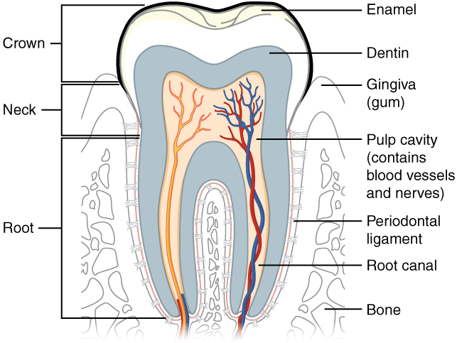
Trace the pulp and root canal to the apical region, then identify the periodontal ligament and supporting bone. Relate these structures to avulsion, apical pathology and extraction. The original uses the spelling “dentin”; cementum is not separately labelled.
Tooth & supporting tissues · OpenStax College · CC BY 3.0. Original image; displayed at a responsive size. Original publication.
Dentine: tubules, odontoblasts and defensive change
Dentine contains a collagen-rich mineralised matrix traversed by tubules extending from the pulpal interface towards enamel or cementum. Odontoblast cell bodies line the pulp; their processes extend into tubules. Tubule density and diameter increase towards the pulp, helping explain why deep dentine is more permeable and why remaining dentine thickness influences pulpal injury.
Primary dentine forms during development, secondary dentine continues more slowly after root formation, and tertiary dentine is produced locally in response to injury. Surviving odontoblasts can form reactionary dentine; replacement odontoblast-like cells can form reparative dentine after greater injury. Dentinal sclerosis reduces tubule patency. These responses can protect the pulp but do not guarantee that an active carious lesion has become harmless.
Oral mucosa: epithelium, basement membrane and lamina propria
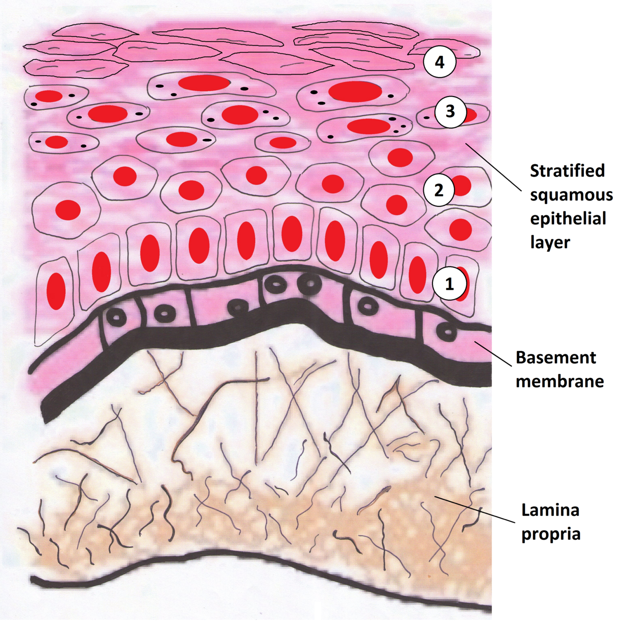
Identify the epithelium, basement membrane and lamina propria. Relate the epithelial–stromal interface to in-situ disease and invasion. This drawing represents a keratinising surface; oral sites have different keratinisation patterns. Use the printed anatomical names for revision.
Oral mucosa: epithelium, basement membrane and lamina propria · Wiki-minor (own work, 2011) · CC BY-SA 3.0. Unchanged published raster preview; displayed responsively. Original available from the source.
Pulp: a vascular connective tissue in a rigid chamber
The pulp contains vessels, nerves, fibroblasts, immune cells and an extracellular matrix. The odontoblast layer borders the predentine, the unmineralised matrix awaiting mineralisation. Vessels and nerves enter mainly through apical foramina, with accessory communications possible. Inflammation within a low-compliance chamber affects local pressure and perfusion; this is a biological process, not a state that can be read directly from pain intensity.
Hydrodynamic movement in exposed patent dentinal tubules contributes to brief dentine sensitivity. Pulpal pain and sensibility testing depend on neural responses, whereas vitality concerns the living vascular supply. A cold response cannot establish the exact histological extent of inflammation. Symptoms, caries depth, restorative history, other tests and imaging must be integrated.
Tooth development: cap, bell and root formation
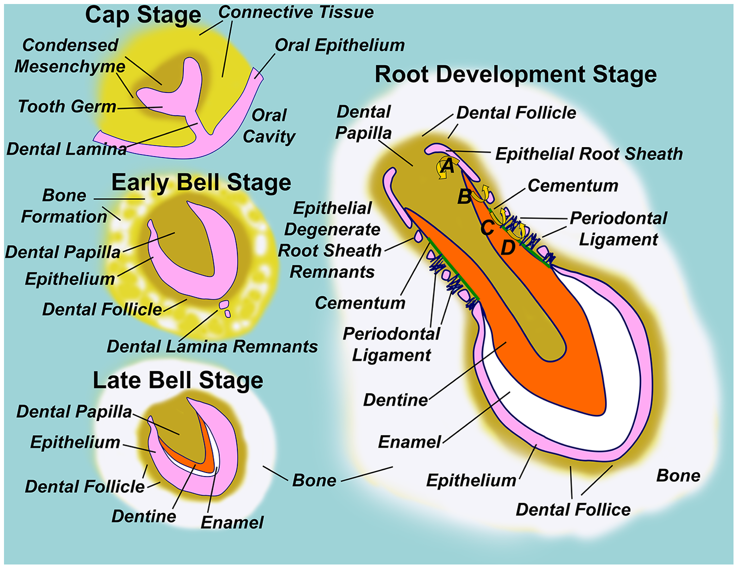
Follow the dental lamina, enamel organ, dental papilla and dental follicle through development. Locate the epithelial root sheath in the root-development panel.
Diagram illustrating normal tooth formation.png · Sarrafpour B, Swain M, Li Q, Zoellner H · CC BY 4.0. None; reproduced without alteration.
Which structure gives rise to dentine and pulp?
The dental papilla. The enamel organ gives rise to enamel; the dental follicle contributes to supporting periodontal tissues.
Cementum, periodontal ligament and alveolar bone
Cementum covers root dentine and provides attachment for periodontal ligament fibres. Acellular cementum is particularly associated with attachment in cervical root regions; cellular cementum is more prominent apically and in furcations. Cementum is normally avascular and differs from bone in its pattern of remodelling. Exposed cementum and root dentine are vulnerable to root caries.
The periodontal ligament is a vascular, innervated connective tissue joining cementum to alveolar bone through collagen fibre insertions. It supports load transfer, proprioception and tissue turnover. Alveolar bone continually remodels through osteoblast and osteoclast activity, which permits orthodontic movement and adaptation. Percussion tenderness therefore concerns supporting tissues and is not a direct test of pulpal vitality.
Extrinsic tongue muscles and surface landmarks
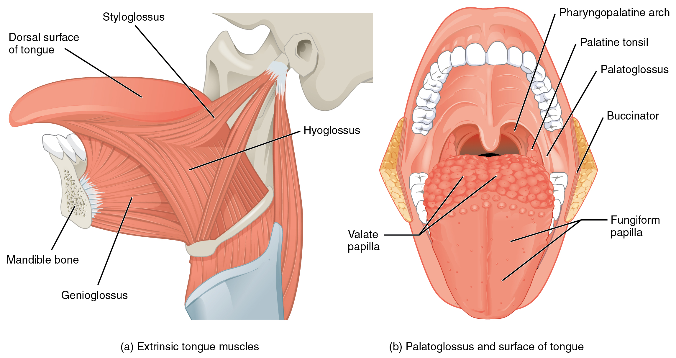
Follow genioglossus from the mandible, hyoglossus from the hyoid and styloglossus from the styloid region. The second view shows palatoglossus and tongue surface landmarks. “Pharyngopalatine arch” is the palatopharyngeal arch; “valate” in the original refers to vallate papillae.
1109 Muscles that Move the Tongue.jpg · OpenStax · CC BY 3.0. None; original image reproduced without alteration.
Which extrinsic tongue muscle is the exception to hypoglossal motor supply?
Palatoglossus, which is supplied through the pharyngeal plexus by the vagus nerve. The other extrinsic tongue muscles receive hypoglossal supply.
Oral mucosa: match the structure to the site
Oral mucosa has stratified squamous epithelium supported by a basement-membrane zone and connective-tissue lamina propria. Basal cells proliferate and differentiate as they move towards the surface. Keratinisation provides mechanical protection at sites exposed to loading; a submucosa containing fat or glands is present in some regions and absent where tissue is tightly bound.
Masticatory mucosa covers gingiva and hard palate and is usually keratinised or parakeratinised. Lining mucosa, including lips, cheeks, floor of mouth, ventral tongue and soft palate, is generally non-keratinised and more flexible. Specialised dorsal tongue mucosa contains papillae; filiform papillae are principally mechanical, while other papillary types are associated with taste buds. Normal regional variation must be recognised before labelling a surface abnormal.
| Tissue | Distinctive structure | Clinical meaning |
|---|---|---|
| Enamel | Highly mineralised, acellular after eruption | Chemical remineralisation is possible; cellular regrowth is not |
| Dentine–pulp complex | Tubules linked to odontoblasts and vascular pulp | Deep injury can affect the pulp without visible exposure |
| Periodontal ligament | Vascular, sensory fibrous attachment | Load, mobility and percussion involve supporting tissues |
| Lining mucosa | Flexible non-keratinised stratified epithelium | Different normal appearance from gingiva or hard palate |
The pulp–dentine interface
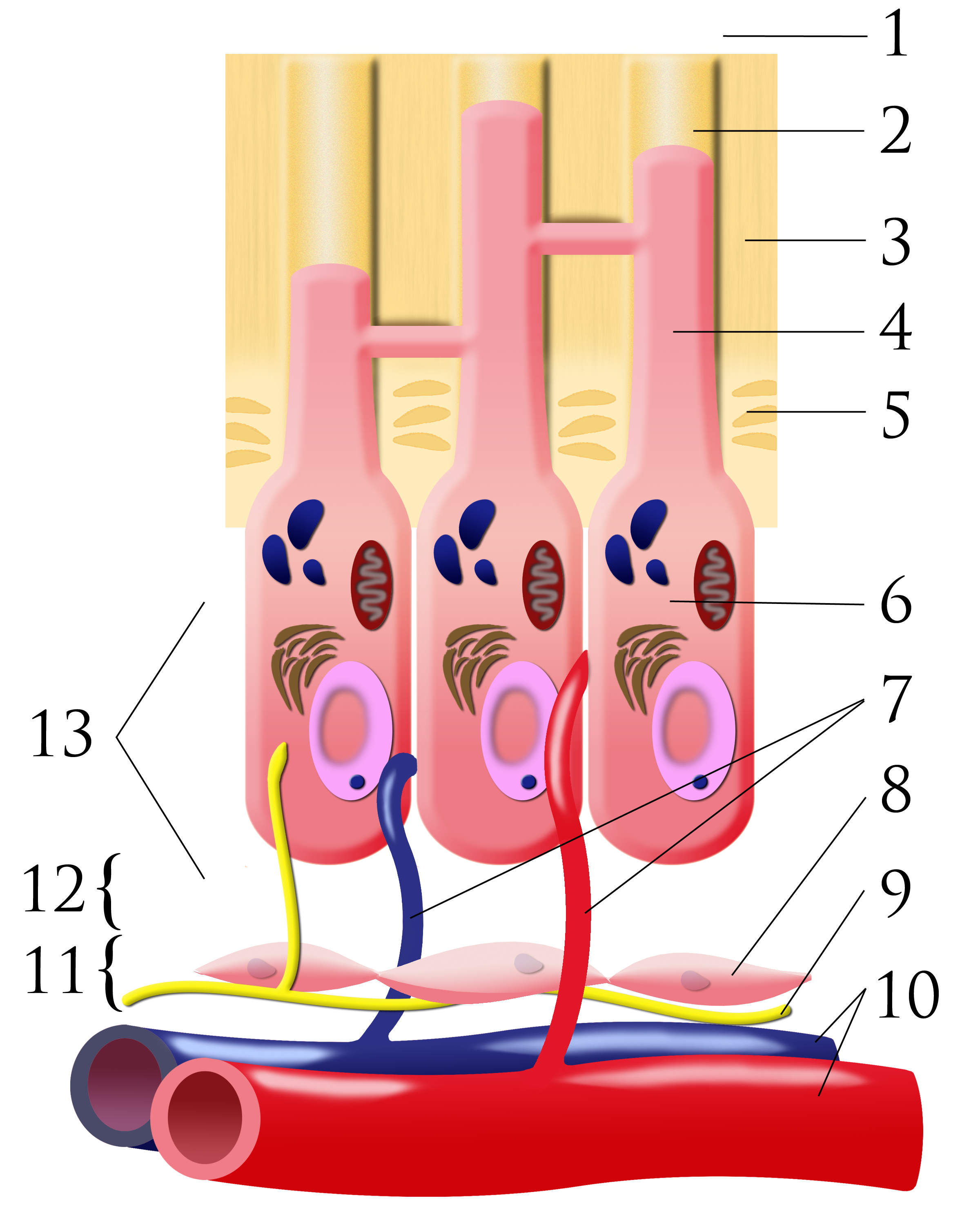
Number key: 1 outer-tooth direction; 2 dentinal tubule; 3 dentine; 4 odontoblastic process; 5 predentine; 6 odontoblast; 7 capillaries; 8 fibroblasts; 9 nerve; 10 artery and vein; 11 cell-rich zone; 12 cell-poor zone; 13 pulp chamber. This is a conceptual schematic, not a scale histological section.
Pulpal dentin junction.png · Ian Furst · CC BY-SA 3.0. None; original image reproduced without alteration.
Which cells form dentine, and where are their cell bodies?
Odontoblasts form dentine. Their cell bodies line the pulp–dentine border, with processes extending into dentinal tubules.
Gingival attachment and the tooth–tissue interface
Oral gingival epithelium faces the mouth, sulcular epithelium lines the sulcus, and junctional epithelium attaches to the tooth through specialised epithelial attachment structures. The junctional epithelium is relatively permeable and permits defence-cell migration. Its normal immune activity does not mean there is no meaningful barrier.
Plaque-related inflammation changes epithelial and connective-tissue relationships. Gingivitis can cause bleeding and swelling without the attachment loss that defines periodontitis. Recession, pocketing and attachment loss must be measured against appropriate landmarks. A deeper sulcus caused by enlargement is not necessarily the same as destruction of the supporting attachment.
Odontoma: dental hard-tissue architecture
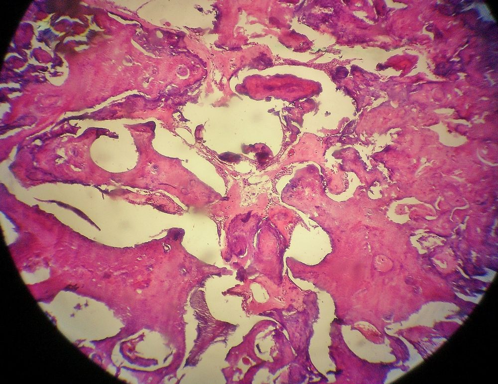
The source identifies an odontoma. Inspect how the dental tissues are organised and compare this tissue view with the atlas’s radiographic odontoma panels, which show different teaching cases. Imaging describes the mineralised pattern and tooth relationships; histology assesses the tissue architecture.
Odontoma.jpg · JXtra · CC BY-SA 3.0. Reproduced without alteration. Labels, arrows and circles, where present, belong to the source image.
Why keep the radiographic and histological descriptions distinct?
They describe different aspects of the lesion. A radiopaque pattern is not itself a microscopic diagnosis, and the specimen must be interpreted with the case information.
Salivary glands and interpreting a tissue specimen
Salivary glands contain secretory acini and ducts. Serous cells produce a watery protein-containing secretion, while mucous cells produce mucins. Parotid tissue is predominantly serous, submandibular mixed with a serous predominance, and sublingual mixed with a mucous predominance. Ducts modify the primary secretion and deliver it to the oral cavity.
Histological interpretation begins with orientation: identify surface epithelium, basement-membrane region, connective tissue and any glands or deeper structures. Inflammation, epithelial dysplasia and invasion are different observations. A small specimen may not represent a whole lesion, so the pathological finding must be reconciled with the clinical site, morphology and sampling method.
A distinction to remember
A positive cold response demonstrates neural responsiveness, not normal histology or measured blood flow. Enamel remineralisation is also different from cellular tissue regeneration.
A cervical root surface gives a brief sharp pain to air that stops when the stimulus stops. How can histology explain that symptom?
Reveal the reasoning
Exposed patent dentinal tubules permit fluid movement that can stimulate pulpal nerves. The pattern is compatible with dentine hypersensitivity, but assess caries, cracks and restoration or pulpal causes before assigning that diagnosis. Persistent spontaneous or lingering pain requires a broader explanation.
Test your recall
Why can early enamel mineral loss reverse even though enamel has no living cells?
Show answer
Mineral ions can diffuse into a non-cavitated porous lesion when the chemical environment favours remineralisation. This repairs mineral balance within existing structure; it does not require ameloblasts or mean that a cavitated defect will grow replacement enamel.