
Caries detection and activity
Detection identifies a lesion; assessment determines its severity, likely activity and clinical consequences. A restorative decision needs all three, together with pulp status and the ability to control the disease.
Quick revision summary
Start with a reproducible visual examination
Examine a clean surface with good illumination and appropriate drying. Remove plaque and debris that hide fissures, proximal margins or cervical lesions. Describe the site, surface integrity, opacity, texture and cavitation using a consistent recording system.
Examine restorations without assuming replacement
Assess an existing restoration’s margins, contact, contour, fracture, wear, retention and effect on the pulp and periodontium. Marginal staining alone does not prove recurrent caries. An accessible local defect may be amenable to finishing, sealing or repair when the remainder is satisfactory.
Record uncertainty and review intelligently
Create a baseline that can be compared: tooth and surface, extent, activity signs, symptoms, image reference and the intended intervention. When monitoring is appropriate, explain what change would trigger treatment and make a specific review plan.
Common mistake
Detection aids and radiographs support a diagnosis; neither replaces examination of activity, surface integrity, pulp and maintainability.
Caries detection and activity
Detection identifies a lesion; assessment determines its severity, likely activity and clinical consequences. A restorative decision needs all three, together with pulp status and the ability to control the disease.
Start with a reproducible visual examination
Examine a clean surface with good illumination and appropriate drying. Remove plaque and debris that hide fissures, proximal margins or cervical lesions. Describe the site, surface integrity, opacity, texture and cavitation using a consistent recording system. An intact enamel lesion, a small surface breakdown and a large dentinal cavity represent different findings even when the patient reports no pain.
Use a gentle tactile examination when it adds information; do not force a sharp explorer into a potentially intact lesion. A stained fissure is not automatically carious, while apparently sound enamel can conceal a proximal lesion. Distinguish a developmental opacity, erosion or an existing restoration defect from caries by distribution, history and the full clinical appearance.
Tooth & supporting tissues
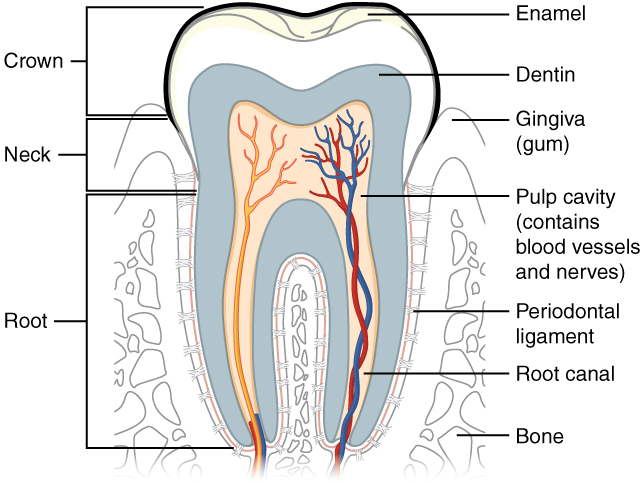
Trace the pulp and root canal to the apical region, then identify the periodontal ligament and supporting bone. Relate these structures to avulsion, apical pathology and extraction. The original uses the spelling “dentin”; cementum is not separately labelled.
Tooth & supporting tissues · OpenStax College · CC BY 3.0. Original image; displayed at a responsive size. Original publication.
Assess activity separately from depth
Activity is inferred from the current lesion and its environment. Matte, rough enamel in a stagnation area with associated plaque is more suggestive of activity; a smooth, shiny surface in a cleansable site may indicate arrest. Exposed root lesions are assessed with texture and accessibility, not simply darkness. Record why the interpretation is reasonable and use serial findings when uncertainty remains.
Depth describes extent of mineral loss, not the rate at which it is occurring. A long-standing deep lesion may be stable, while a shallow lesion can be active in a high-risk patient. The same patient can have both. Risk assessment guides the strength of prevention and review but cannot substitute for examining each relevant surface.
Use imaging to answer a defined question
Justified bitewings can reveal proximal lesions, estimate dentinal extent and assess areas concealed from direct examination. Compare previous images with attention to projection and exposure differences. A radiograph is a two-dimensional representation of mineral loss and may not match the true clinical depth. It does not demonstrate whether an accessible surface is cavitated or establish current activity by itself.
Periapical imaging may be indicated when symptoms, depth or examination raise a pulpal or apical question. Supplementary methods such as transillumination can add information at selected sites, but an instrument reading is not an independent treatment instruction. Understand false positives and the effect of stain, restorations and access before interpreting a detection aid.
Proximal caries in primary molars
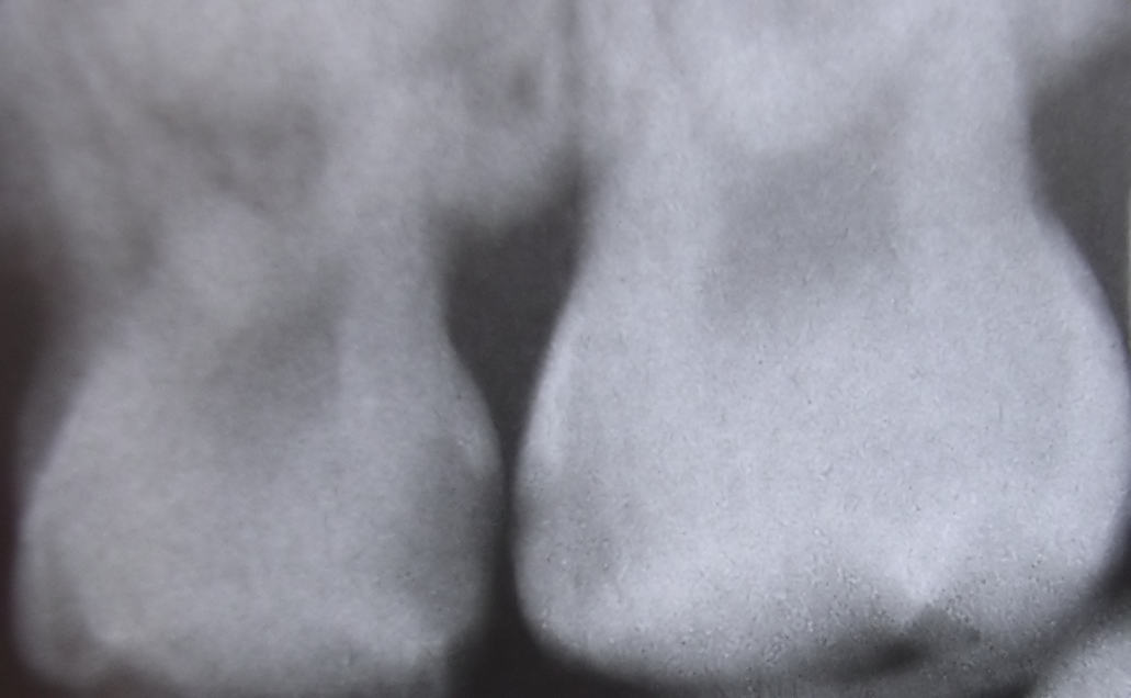
The source identifies interproximal caries between upper primary molars. Describe the radiolucent areas beneath the contact, then assess activity and cavitation clinically.
Interproximaldecayfiltered08-16-2006.jpg · Dozenist · CC BY-SA 3.0. Source author’s Neat Image processing retained. No additional image changes.
Examine restorations without assuming replacement
Assess an existing restoration’s margins, contact, contour, fracture, wear, retention and effect on the pulp and periodontium. Marginal staining alone does not prove recurrent caries. An accessible local defect may be amenable to finishing, sealing or repair when the remainder is satisfactory. Distinguish a superficial imperfection from a disease process extending beyond the visible margin.
Imaging and clinical findings should agree with the proposed problem. A radiolucent lining or projection artefact can be mistaken for decay. If caries is suspected, describe where and why, and consider whether partial removal or exploration will alter management. Replacing the whole restoration irreversibly removes more tissue and should have a clear indication.
Impacted third molar beside distal second-molar caries
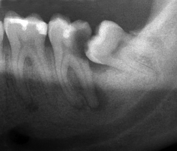
The source identifies caries on the second molar beside an impacted lower third molar. Describe the tooth positions and adjacent surface before deciding the clinical significance.
Wisdom tooth causing dental caries.png · Coronation Dental Specialty Group · CC BY-SA 3.0. Image reproduced without alteration; original marks and any embedded credits retained.
Convert the findings into options
A non-cavitated, maintainable lesion may be managed through prevention, sealing or a selected micro-invasive option with monitoring. An active cavity that cannot be cleaned may need restoration to create a sealed, maintainable surface. Structural damage, symptoms, progression and patient priorities also matter. There is no single radiographic line that makes the decision for every tooth.
Before treating a deep lesion, assess symptoms, sensibility, percussion, restorability and the ability to isolate. Discuss pulp exposure and possible vital pulp or root canal treatment if the findings change. The aim is not simply to detect more lesions but to choose interventions that improve tooth survival while avoiding unnecessary loss of tissue.
| Finding | What it supports | What remains to assess |
|---|---|---|
| Intact early lesion | Potential non-operative control | Activity, risk and review feasibility |
| Uncleanable active cavity | Possible restorative indication | Pulp, extent and restoration design |
| Stained margin | A colour observation | Caries, seal, fracture and symptoms |
| Deep radiolucency | Potential pulp risk | Clinical surface and pulpal diagnosis |
Record uncertainty and review intelligently
Create a baseline that can be compared: tooth and surface, extent, activity signs, symptoms, image reference and the intended intervention. When monitoring is appropriate, explain what change would trigger treatment and make a specific review plan. A note that says watch does not reveal whether the clinician intends to observe activity, cavitation or a restoration defect.
At review, compare like with like and reassess the patient’s exposures and prevention. Progression despite a genuinely implemented plan may justify escalation; persistent colour without activity may not. New pain requires diagnostic reassessment rather than waiting for a planned image interval. A coherent record links the observation, interpretation, choice and subsequent response.
A distinction to remember
Detection aids and radiographs support a diagnosis; neither replaces examination of activity, surface integrity, pulp and maintainability.
A proximal radiolucency reaches outer dentine, but the surface is intact and previous images are stable. Must it be restored?
Reveal the reasoning
Not automatically. Consider lesion activity, cavitation, risk, cleansability and confidence in the image comparison. A preventive or selected micro-invasive approach with documented monitoring may be appropriate. Radiographic depth alone does not establish that operative treatment offers the best balance.
Test your recall
What should be recorded when a lesion is detected?
Show answer
Record tooth and surface, severity, surface integrity, activity signs, symptoms and relevant image findings. Use a consistent system so another clinician can compare the site. A binary caries-present entry cannot explain whether prevention, monitoring or restoration was selected.