
Bitewings and periapicals
A technically limited image can falsely reassure or create apparent disease.
Quick revision summary
Choose and assess the intraoral projection
Assess identification, coverage, positioning, angulation, sharpness and exposure before interpreting an intraoral image. Proximal overlap may obscure caries; a missed apex or distorted root may defeat the diagnostic aim.
Use the view that answers the question
Bitewings are useful for proximal caries and crestal bone assessment when contacts and the relevant anatomy are adequately shown. A periapical view includes the roots and apical tissues and can support endodontic or local bone assessment.
Connect the image to the clinical surface
Radiographic caries depth is an estimate of mineral loss. It does not demonstrate cavitation, activity or whether a lesion is accessible to plaque control.
Common mistake
Image enhancement cannot repair missing coverage or remove the diagnostic effect of overlapping anatomy.
Bitewings and periapicals: quality before interpretation
A technically limited image can falsely reassure or create apparent disease.
Choose and assess the intraoral projection
Assess identification, coverage, positioning, angulation, sharpness and exposure before interpreting an intraoral image. Proximal overlap may obscure caries; a missed apex or distorted root may defeat the diagnostic aim. The paralleling technique supports reproducibility where feasible. Alternative approaches may be needed for anatomy or tolerance, with their limitations recorded.
Interpret proximal surfaces, restorations, pulp spaces, roots, periodontal ligament, lamina dura and supporting bone in an orderly sequence. Compare prior suitable images when progression is the question. A radiolucency needs clinical correlation and awareness of normal anatomy or artefacts. Caries extent on an image does not establish cavitation or activity. Repeat an exposure only when the missing information is necessary and a corrected technique is feasible.
Nasopalatine duct cyst: an anterior maxillary example
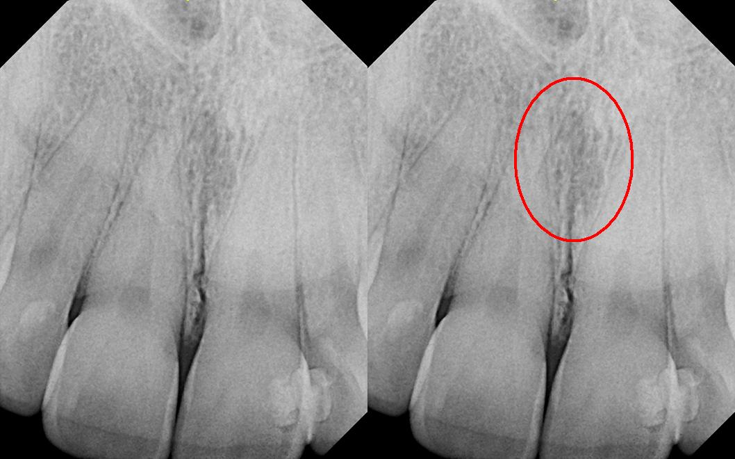
The right panel repeats the image with the source’s red circle highlighting the midline region. The source explicitly corrects its filename: this is reported as a nasopalatine duct cyst, not a nasolabial cyst. Consider the incisive canal and adjacent incisors, including pulp tests. The subtle appearance illustrates why a labelled example is not a stand-alone diagnostic rule.
Nasolabial_duct_cyst.JPG · DRosenbach · CC BY-SA 3.0. Source image reproduced without alteration; any arrows or circles are part of the original.
Why assess the adjacent incisors before treating a midline anterior maxillary radiolucency as endodontic disease?
A nasopalatine duct cyst or normal incisive-canal anatomy may overlap the incisor roots. Clinical examination, pulp tests and appropriate imaging help avoid unnecessary endodontic treatment of otherwise healthy teeth.
Key distinctions in practice
| Finding or option | What it means | Clinical consequence |
|---|---|---|
| Overlap | Contact projection obscures surfaces | May limit proximal caries assessment. |
| Apex omitted | The periapical region is not captured | Decide whether the diagnostic question remains unanswered. |
| Elongation / foreshortening | Projection changes apparent form | Avoid treating distorted dimensions as anatomical truth. |
| Reproducible view | Comparable geometry over time | Helps assess genuine change rather than positioning differences. |
Use the view that answers the question
Bitewings are useful for proximal caries and crestal bone assessment when contacts and the relevant anatomy are adequately shown. A periapical view includes the roots and apical tissues and can support endodontic or local bone assessment. Neither is an automatic substitute for the other. Select the examination from the clinical question and available previous images.
A holder and a reproducible paralleling technique can improve geometry where feasible. Check the coverage before interpreting: a cut-off apex cannot answer an apical question, while overlapping contacts may hide proximal lesions. Digital enhancement can improve viewing of existing information but cannot reconstruct anatomy that was not captured.
Periodontal bone loss on a periapical radiograph
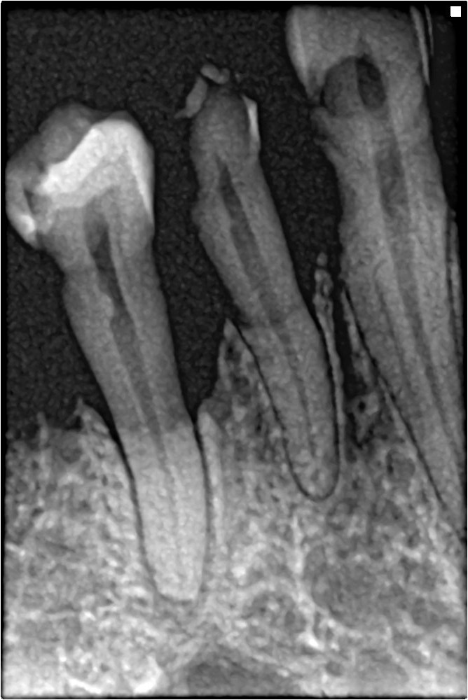
Trace the reduced bone height and irregular contour between the teeth. Correlate with full periodontal charting; this single image cannot establish the patient’s complete stage and grade.
Bone loss in periapical xray.jpg · Shaimaa Abdellatif · CC BY-SA 4.0. Image reproduced without alteration; original marks and any embedded credits retained.
Connect the image to the clinical surface
Radiographic caries depth is an estimate of mineral loss. It does not demonstrate cavitation, activity or whether a lesion is accessible to plaque control. Combine the image with clinical examination and disease risk when planning prevention or operative treatment. Likewise, bone levels need correlation with probing and the distribution of disease.
When reviewing serial images, consider whether projection and exposure are sufficiently comparable. Apparent change can arise from geometry or image quality. If the image is inadequate, state which question remains unanswered. Repeat only when the additional information is justified and the cause of the error can be addressed, rather than repeating simply to obtain a more attractive picture.
Proximal caries in primary molars
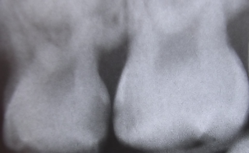
The source identifies interproximal caries between upper primary molars. Describe the radiolucent areas beneath the contact, then assess activity and cavitation clinically.
Interproximaldecayfiltered08-16-2006.jpg · Dozenist · CC BY-SA 3.0. Source author’s Neat Image processing retained. No additional image changes.
A distinction to remember
Image enhancement cannot repair missing coverage or remove the diagnostic effect of overlapping anatomy.
A bitewing has overlapping proximal contacts. Can it reliably exclude early caries at those surfaces?
Reveal the reasoning
No. Overlap limits assessment. Record the limitation and decide whether a justified corrected view or other assessment is needed.
Guidance to read alongside this chapter
Test your recall
Why might overlapping contacts limit caries assessment?
Show answer
The proximal surfaces are obscured.