
Salivary gland disease
Salivary symptoms fall into three overlapping patterns: a flow problem, inflammatory enlargement and a persistent mass. Their investigation differs. Distinguish the sensation of a dry mouth from measured reduction in salivary output, and distinguish an intermittently blocked duct from a lump that remains between meals.
THE ANATOMY LAB / INTERACTIVE 3DExplore the salivary gland relationships in 3D↗Quick revision summary
Follow the timing
Pain and swelling with meals suggest obstructed salivary flow. A persistent lump needs its own assessment, even if painless. Record the gland, duct findings, facial movement and any cervical lymphadenopathy.
Separate dryness from its cause
Xerostomia is a symptom; salivary hypofunction is a measured finding. Medicines, dehydration, radiotherapy and systemic disease are possible contributors. Dry mouth alone does not establish Sjögren disease or justify stopping prescribed medication.
Protect function while investigating
Prevention, hydration and symptom relief support patients while the cause is investigated. Recurrent obstruction may suit gland-preserving treatment. Rapid progression, facial weakness, systemic illness or swallowing and airway symptoms require a different urgency.
Common mistake
Meal-related swelling suggests obstruction; a painless persistent mass is not thereby benign. Facial weakness or a rapidly progressive neck swelling must not be managed as routine dry mouth.
Salivary gland disease: obstruction, dry mouth and persistent masses
Salivary symptoms fall into three overlapping patterns: a flow problem, inflammatory enlargement and a persistent mass. Their investigation differs. Distinguish the sensation of a dry mouth from measured reduction in salivary output, and distinguish an intermittently blocked duct from a lump that remains between meals.
Use anatomy to localise the complaint
The parotid lies around the posterior mandibular ramus and drains through Stensen’s duct opposite the upper second molar. The submandibular gland has superficial and deep parts related to mylohyoid; Wharton’s duct opens beside the lingual frenulum. Sublingual and minor glands contribute saliva through multiple small ducts. The facial nerve traverses the parotid, whereas the lingual and hypoglossal nerves are relevant to submandibular surgery.
Ask the patient to point to the swelling and describe whether it enlarges during eating, resolves afterwards or remains continuously present. A mandibular dental abscess, lymph node or soft-tissue lesion can resemble a gland problem. Examine both sides rather than assuming that every swelling near the jaw angle arises in the parotid.
Major salivary glands & ducts
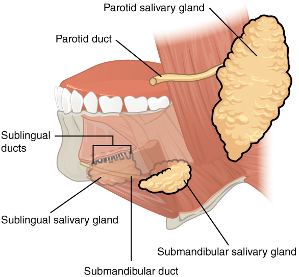
Name each gland and its duct opening. Relate duct obstruction to meal-related swelling and the sublingual gland to a ranula. This overview does not depict every lingual-nerve or facial-nerve relationship.
Major salivary glands & ducts · OpenStax College · CC BY 3.0. Original image; displayed at a responsive size. Original publication.
Recognise obstruction and acute inflammation
Salivary stimulation against a stone or stricture can produce meal-related pain and enlargement. Palpate the floor of the mouth gently, inspect duct openings and assess the character and amount of expressed saliva. A stone may be palpable, but absence of a visible stone does not exclude obstruction farther along the duct or within the gland.
Acute bacterial sialadenitis can cause tenderness, swelling and purulent discharge, particularly with impaired flow or dehydration. Evaluate systemic illness and any spreading neck or floor-of-mouth process. Infection and obstruction may coexist; repeated antibiotic courses without addressing reduced flow or the obstructive cause can leave the underlying problem unresolved.
Coronal sublingual and submandibular spaces with normal MRI correlation
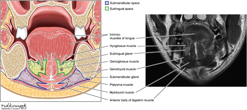
Identify the mylohyoid and compare the sublingual and submandibular compartments in the schematic and normal MRI. Relate the compartments to the tooth-apex relationships discussed in this section.
Coronal sublingual and submandibular spaces with normal MRI correlation · Swapnil Patel and Alok A. Bhatt (2018); illustration credited within the figure to Nadezhda D. Kiriyak / University of Rochester · CC BY 4.0. Unchanged published image; displayed responsively. Original publication.
Investigate a gland or duct with a defined question
Ultrasound can assess many superficial gland lesions, ducts and stones and can guide sampling. CT may help localise stones or define acute deep-space complications; MRI is useful for selected soft-tissue or tumour questions. The modality depends on site, suspected disease and the management decision. Not every stone is visible on a routine dental radiograph.
Selected obstruction can be treated with gland-preserving approaches, including sialendoscopy or directed stone removal. Gland excision is a separate decision with nerve and other operative risks. Do not try to cut out a floor-of-mouth swelling without establishing its anatomy and diagnosis. Hydration and appropriate conservative advice should accompany a definitive assessment plan.
Major salivary glands
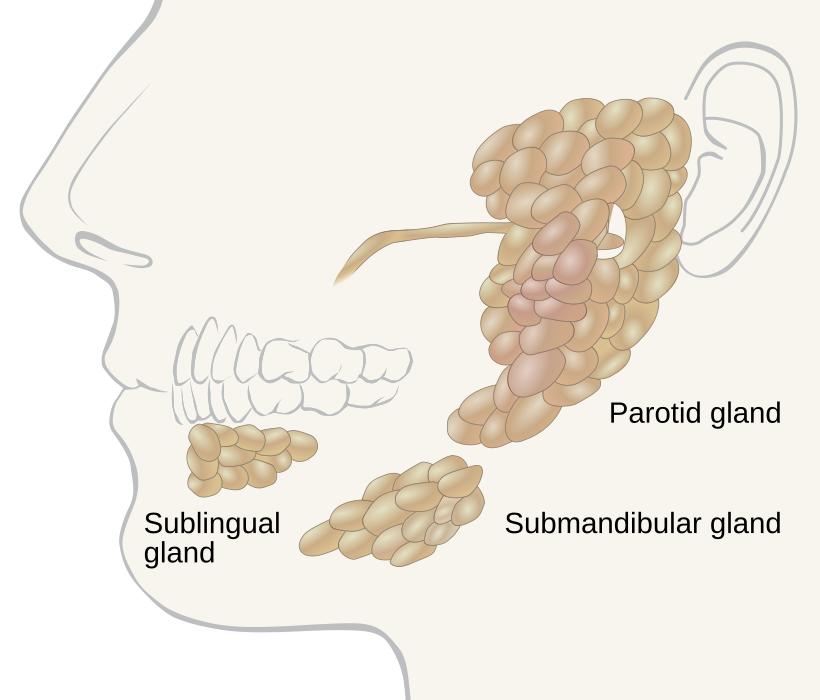
Locate the parotid, submandibular and sublingual glands. Use the gland distribution to relate swelling to the history; the diagram alone does not establish the cause of a mass.
Salivary glands labeled.svg · Goran tek-en, following request by and knowledge from Tom (LT) · CC BY-SA 4.0. None; reproduced without alteration.
Which major gland sits in the anterior floor of the mouth?
The sublingual gland. The submandibular duct also opens into the anterior floor of the mouth, near the lingual frenulum.
Assess dry mouth as a symptom with consequences
Ask about onset, variation, difficulty eating dry food, nocturnal drinking, altered taste and soreness. Review all medicines, including over-the-counter preparations, and relevant conditions or cancer treatment. Examine mucosal moisture, pooled saliva, saliva consistency, candidosis, cervical/root caries and denture tolerance. Symptoms and flow measurements can differ, so one should not automatically substitute for the other.
Management combines treatment of reversible contributors with protection of the teeth and mucosa. Consider water, suitable saliva substitutes or safe salivary stimulation where residual function exists. Avoid frequent sugary or acidic drinks used to relieve dryness. Agree fluoride-based prevention, dietary support and review according to the actual caries risk. Medication changes require the prescriber’s involvement.
Submandibular duct stone at the opening
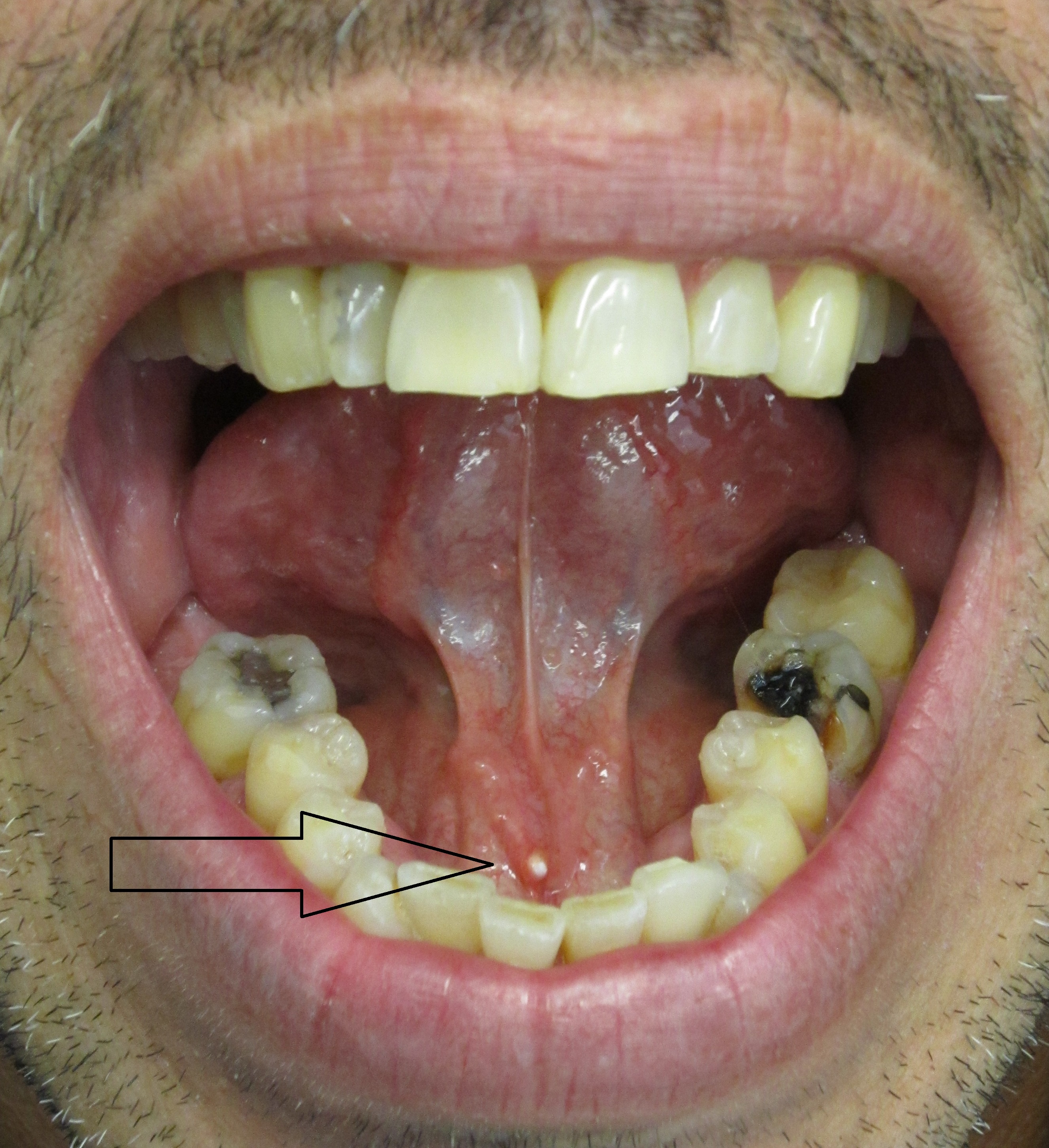
Use the original arrow to locate the pale stone at the submandibular duct opening. Relate the position to the lingual frenulum. Ask about meal-related swelling and assess the gland, duct and saliva. A visible distal calculus does not exclude another obstruction or associated infection elsewhere in the system.
Salivary stone in submandibular salivary duct.jpg · James Heilman, MD · CC BY-SA 3.0. Reproduced without alteration. Labels, arrows and circles, where present, belong to the source image.
Why can gland swelling become more noticeable around meals?
Salivary stimulation increases flow against an obstruction, which can produce swelling and pain. Correlate the timing with examination rather than diagnosing every meal-related swelling as a stone.
Consider Sjögren disease without overdiagnosing it
Persistent oral and ocular dryness, recurrent gland enlargement and systemic features can suggest Sjögren disease, an immune-mediated disorder. Assessment may combine history, examination, blood tests, ocular testing, salivary flow and specialist-selected gland biopsy. No single symptom, positive antibody or negative antibody result establishes or excludes every case.
Coordinate dental care with oral medicine and the relevant medical team. Ask about fatigue, joint or other systemic symptoms and changes in gland swelling. A new persistent or asymmetric enlargement requires review rather than being attributed automatically to known Sjögren disease. Long-term care addresses oral complications as well as the systemic diagnosis.
Mucocele: low-power tissue appearance
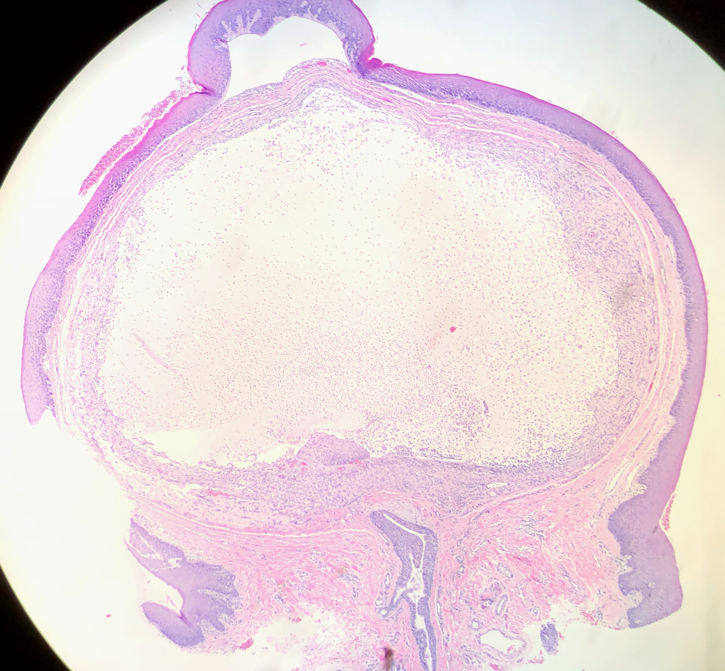
The source identifies a lower-lip mucocele. Orient yourself at the surface epithelium, then find the large pale mucin-rich area beneath it and the surrounding tissue reaction. Distinguishing mucous extravasation from a duct-lined retention lesion requires attention to the complete specimen and report.
Mucocele of Lower Lip (49425670122).jpg · Ed Uthman · CC BY 2.0. Reproduced without alteration. Labels, arrows and circles, where present, belong to the source image.
Which tissue relationship is important when interpreting a mucocele specimen?
The relationship of mucin, the surrounding reaction and any duct or epithelial lining. A cavity-like space alone does not establish that it is a true epithelial-lined cyst.
Treat persistent masses as a different diagnostic problem
Benign and malignant tumours can present as painless salivary lumps. Growth, fixation, pain, facial weakness, skin change or cervical nodes increase concern, but their absence does not establish benignity. Record facial movement when assessing a parotid mass and arrange an appropriate specialist pathway.
Imaging and needle sampling are planned around the lesion and possible definitive treatment. Avoid an unplanned open biopsy of a major gland mass. Mucoceles often follow minor-gland trauma, while a ranula is associated with the sublingual region; a plunging ranula can extend into the neck. These diagnoses still need anatomical confirmation and a treatment plan appropriate to recurrence and extent.
A distinction to remember
Meal-related swelling suggests obstruction; a painless persistent mass is not thereby benign. Facial weakness or a rapidly progressive neck swelling must not be managed as routine dry mouth.
A patient has swelling beneath the jaw during meals that settles between episodes. What mechanism should lead the differential?
Reveal the reasoning
Obstructed salivary outflow, such as a submandibular duct stone or stricture, should lead the differential. Examine the gland and duct and look for infection, then select imaging or specialist assessment to localise the problem. The pattern supports obstruction but does not identify its exact cause.
Test your recall
Where do the parotid and submandibular ducts open?
Show answer
Stensen’s duct opens on the buccal mucosa opposite the upper second molar. Wharton’s duct opens at the sublingual caruncle beside the lingual frenulum. Knowing these sites helps examination of flow and discharge, while remembering that deeper obstruction may not be visible.