
Candidosis and infection
Candida can be carried normally in the mouth. Candidosis is clinical infection in a susceptible local or systemic environment, not merely a positive culture. Recognise the different appearances, treat contributing factors and check interactions before choosing antifungal treatment, while keeping persistent atypical lesions under review.
Quick revision summary
Distinguish carriage from disease
Candida species can inhabit the oral environment without symptoms. A diagnosis of infection should fit the appearance, symptoms and predisposing context. Discomfort, altered taste and erythema can have other causes, so a positive swab must be interpreted rather than treated as proof that every symptom is fungal.
Differentiate other infectious and inflammatory disease
HSV can produce vesicles and ulcers, especially widespread gingivostomatitis in primary infection; recurrence and immune suppression alter presentation. VZV can follow a unilateral neurological distribution and associated eye involvement may be urgent.
Review the tissue and prevent recurrence
Assess symptoms and the original lesions after treatment. A positive culture becoming negative is not the only outcome, and repeated cultures are not routinely a substitute for examination. Persistent plaques, focal erosion or induration can require biopsy or another diagnostic pathway.
Common mistake
Topical miconazole can interact significantly with warfarin. A positive Candida swab does not prove that a persistent non-wiping lesion is simply thrush.
Candidosis and oral infection: diagnose the pattern and cause
Candida can be carried normally in the mouth. Candidosis is clinical infection in a susceptible local or systemic environment, not merely a positive culture. Recognise the different appearances, treat contributing factors and check interactions before choosing antifungal treatment, while keeping persistent atypical lesions under review.
Distinguish carriage from disease
Candida species can inhabit the oral environment without symptoms. A diagnosis of infection should fit the appearance, symptoms and predisposing context. Discomfort, altered taste and erythema can have other causes, so a positive swab must be interpreted rather than treated as proof that every symptom is fungal.
A pseudomembranous pattern has removable white plaques over an erythematous surface. An erythematous pattern may involve red depapillated or sore mucosa without obvious white plaques. Document the original distribution and wipe behaviour gently, avoiding aggressive scraping that injures tissue or confuses the baseline appearance.
Oral candidosis: pseudomembranous plaques
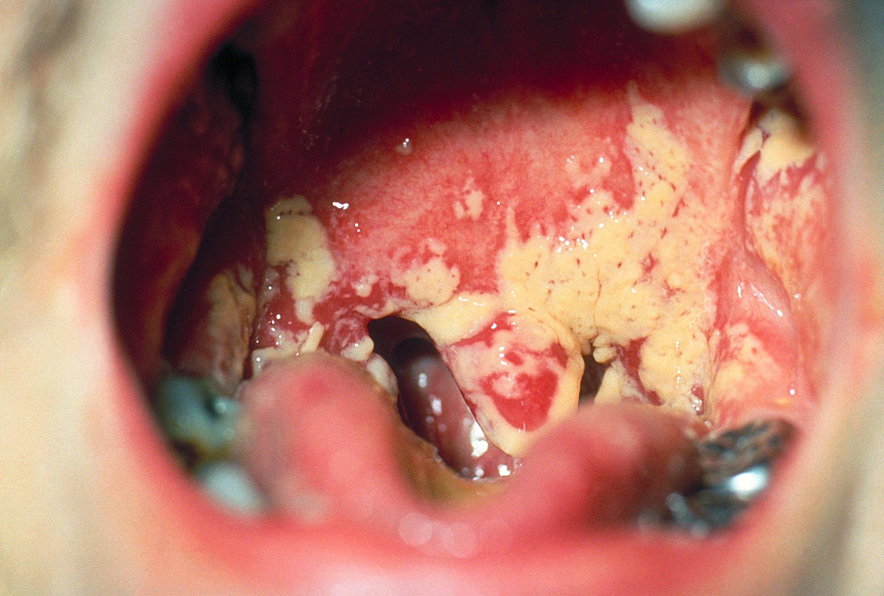
The CDC source identifies oral candidosis. Note the cream-coloured plaques over erythematous mucosa. Pseudomembranous candidosis often has removable deposits, but a static photograph cannot demonstrate that behaviour. Correlate with examination and factors such as medicines, denture use, dry mouth and immunosuppression. The legacy filename includes “Aphthae”; this is not an aphthous-ulcer example.
Oral_thrush_Aphthae_Candida_albicans._PHIL_1217_lores.jpg · Centers for Disease Control and Prevention (CDC), PHIL 1217 · Public domain (US federal government). Source image reproduced without alteration; any arrows or circles are part of the original.
What bedside observation helps distinguish pseudomembranous candidosis from a fixed white plaque?
Whether the deposit can be gently removed, often exposing erythematous mucosa. Interpret this with the history and examination; not every white lesion is candidosis.
Recognise appliance and lip-corner patterns
Denture-associated stomatitis commonly affects the mucosa beneath an appliance and may be relatively symptom-free. Appliance biofilm, prolonged wear, fit, trauma and the local environment interact. Treating the mucosa while ignoring the denture can allow persistence or recurrence.
Angular cheilitis affects the lip commissures and can involve Candida, bacteria, saliva pooling, local irritation or systemic factors. Reduced vertical support and relevant deficiencies may contribute in selected cases. The appearance is not a universal indication for a systemic azole, and a unilateral persistent atypical lesion requires its own assessment.
Angular cheilitis: commissural inflammation
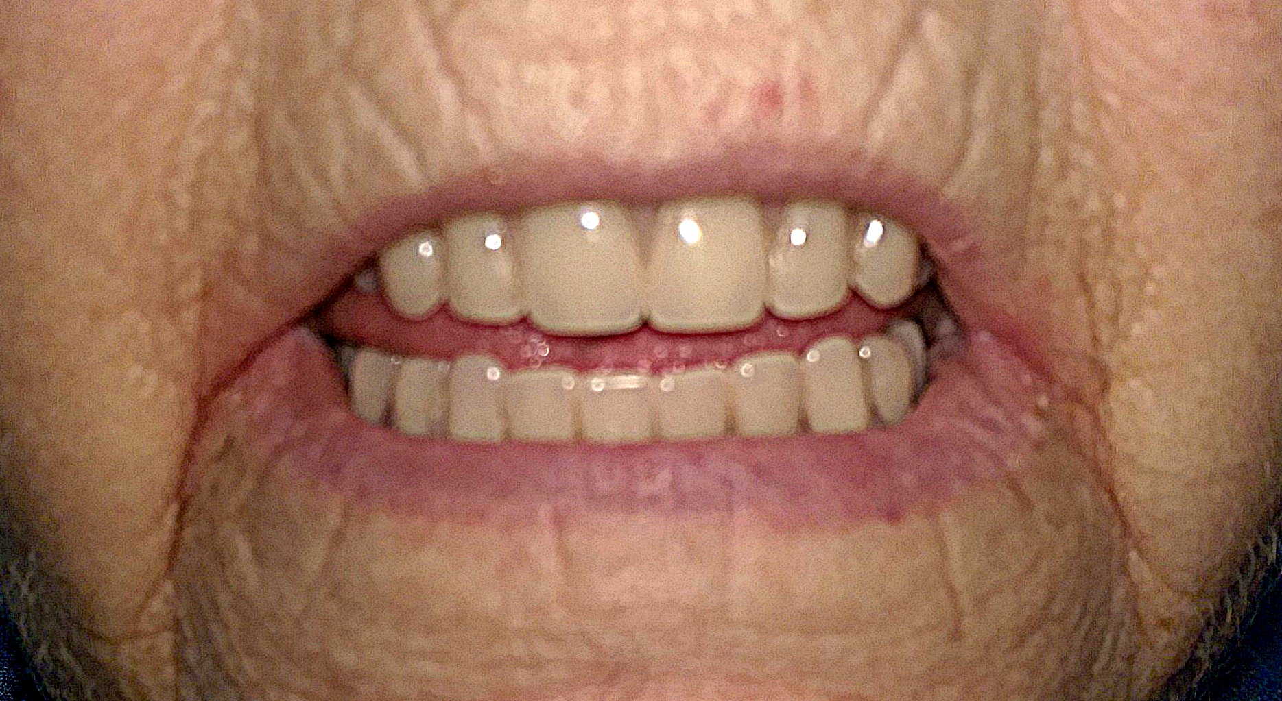
Inspect the mouth corners for redness and fissuring. The source records angular cheilitis in a person with full dentures and deep commissural folds. Assess local moisture, denture factors and relevant medical or nutritional contributors. The visible pattern does not identify Candida or another organism by itself.
Angular cheilitis.jpg · Matthew Ferguson 57 · CC BY-SA 3.0. Reproduced without alteration. Labels, arrows and circles, where present, belong to the source image.
Does the appearance alone establish that Candida is the cause?
No. Angular cheilitis can have several contributing factors. The history, examination and response to appropriate measures guide whether further investigation is needed.
Find the predisposing conditions
Ask about recent broad-spectrum antibiotics, inhaled or systemic corticosteroids, immune-modifying medicines, diabetes, dry mouth, nutritional problems and smoking. Examine denture hygiene and wearing habits, salivary function and mucosal trauma. Age and immune context influence the differential and urgency.
For inhaled corticosteroids, review mouth rinsing and correct technique, with spacer advice where relevant through the appropriate clinician. Do not tell patients to stop necessary respiratory treatment. Persistent or recurrent candidosis without an obvious explanation can justify medical or specialist assessment rather than repeated treatment alone.
Median rhomboid glossitis: central papillary change
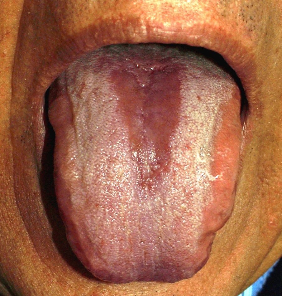
The source identifies median rhomboid glossitis. Locate the relatively smooth red midline area and compare it with the surrounding papillary surface. Describe the site and shape before interpreting the cause. A photograph does not demonstrate fungal organisms, and treatment decisions depend on symptoms and the wider assessment.
Glossitis.jpg · Klaus D. Peter, Wiehl, Germany · CC BY 3.0 DE. Reproduced without alteration. Labels, arrows and circles, where present, belong to the source image.
How does its distribution differ from the geographic-tongue example?
This example has a more localised central area. Geographic tongue shows an irregular map-like pattern whose distribution may change over time.
Differentiate other infectious and inflammatory disease
HSV can produce vesicles and ulcers, especially widespread gingivostomatitis in primary infection; recurrence and immune suppression alter presentation. VZV can follow a unilateral neurological distribution and associated eye involvement may be urgent. These are different mechanisms from fungal plaques and require their own assessment.
Lichen planus, leukoplakia, nutritional glossitis, geographic tongue, chemical injury and malignancy can mimic parts of candidosis. Chronic non-wiping or focal indurated disease should not be explained away by a fungal result. Infection can coexist with an underlying lesion, so partial improvement requires careful re-examination.
Select local measures and antifungals safely
Improve appliance hygiene using methods compatible with the material and give suitable periods out of the mouth. Address trauma, dryness and relevant oral hygiene. These measures are part of treatment, not an optional extra after a prescription. Provide symptom support where eating or brushing is difficult.
Current SDCEP guidance distinguishes mild/localised from extensive or severe infection and directs topical or systemic treatment accordingly. Check the current regimen, formulation, age suitability, medical conditions and interactions. Topical oral miconazole is systemically absorbed enough to cause important interactions; “topical” does not guarantee pharmacological safety.
Check interactions and escalation needs
Miconazole and fluconazole have important interactions, including with warfarin and some statins. Use the current BNF and SDCEP advice rather than adding an azole without a medicine check. Renal or hepatic disease and other therapies can affect systemic antifungal decisions.
Immunocompromised patients with severe infection, systemic illness or swallowing problems may need medical assessment. Dysphagia or odynophagia should raise concern beyond uncomplicated local thrush. If appropriate treatment fails, reconsider the diagnosis, contributing environment, adherence and possible organism or resistance issues, obtaining specialist advice where indicated.
Review the tissue and prevent recurrence
Assess symptoms and the original lesions after treatment. A positive culture becoming negative is not the only outcome, and repeated cultures are not routinely a substitute for examination. Persistent plaques, focal erosion or induration can require biopsy or another diagnostic pathway.
Explain that recurrence may reflect an ongoing predisposing factor rather than an inadequate drug dose. Support appliance care, inhaler technique, preventive dentistry and treatment of the relevant systemic problem. Document follow-up and the escalation plan, especially when the initial presentation was extensive, unexplained or in an immunocompromised patient.
A distinction to remember
Topical miconazole can interact significantly with warfarin. A positive Candida swab does not prove that a persistent non-wiping lesion is simply thrush.
A warfarin-treated patient presents with a sore mouth and removable plaques. What prescribing issue must be checked?
Reveal the reasoning
Assess whether candidosis is clinically supported and check antifungal interactions before treatment. Oral miconazole and fluconazole can interact significantly with warfarin despite miconazole being used topically. Use current SDCEP/BNF advice and an appropriate alternative or coordinated plan rather than routine azole prescribing.
Test your recall
Why can Candida be isolated without a diagnosis of candidosis?
Show answer
It can be part of oral carriage without causing clinical disease. Interpret a result alongside symptoms, lesion appearance and predisposing factors. Treating a swab alone can overlook another diagnosis, particularly when a focal persistent or non-wiping lesion does not fit ordinary thrush.