
Odontogenic infection
An odontogenic infection can range from a local collection to deep-space spread with airway compromise or sepsis. The key decisions are where the source is, how far the infection has spread and how unwell the person is. Antibiotics support selected cases; they do not replace drainage or definitive dental care.
Quick revision summary
Understand the local disease process
Pulpal necrosis and periodontal infection can allow mixed oral organisms to spread through the periapical tissues or periodontal spaces. Inflammation may be localised, diffuse cellulitis or a collection. A fluctuant swelling suggests pus but its absence does not exclude a significant infection, particularly in deep tissues.
Recognise sepsis without reducing it to a temperature
Sepsis is life-threatening organ dysfunction caused by a dysregulated response to infection. Confusion, poor perfusion, respiratory compromise and reduced urine output can be more important than the size of the facial swelling.
Prevent recurrence and communicate the safety net
Explain the cause, what has been drained or removed and any remaining definitive treatment. Check analgesia, hydration and ability to obtain care. Give explicit warning signs and emergency contacts. Where emergency transfer is needed, communicate observations, airway concerns, progression, comorbidity, allergies and medicines already given.
Common mistake
A normal temperature and a small visible swelling do not rule out serious disease. Antibiotics cannot compensate for an unprotected airway or an untreated source.
Odontogenic infection: source, spaces and sepsis
An odontogenic infection can range from a local collection to deep-space spread with airway compromise or sepsis. The key decisions are where the source is, how far the infection has spread and how unwell the person is. Antibiotics support selected cases; they do not replace drainage or definitive dental care.
Understand the local disease process
Pulpal necrosis and periodontal infection can allow mixed oral organisms to spread through the periapical tissues or periodontal spaces. Inflammation may be localised, diffuse cellulitis or a collection. A fluctuant swelling suggests pus but its absence does not exclude a significant infection, particularly in deep tissues.
Identify the source through history, caries and periodontal assessment, tenderness, relevant sensibility testing and imaging. A draining sinus may temporarily reduce pain while the source persists. Distinguish dental infection from salivary disease, infected cyst, mucosal lesions and other facial or neck swellings.
Mandibular landmarks
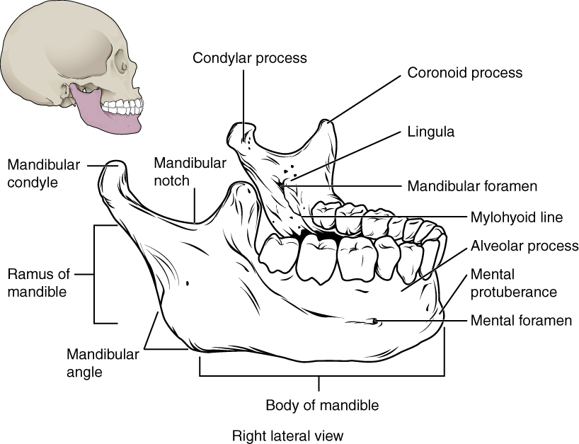
Locate the mental and mandibular foramina, lingula and mylohyoid line. The mandibular foramen, lingula and mylohyoid line are medial landmarks; this schematic brings several surfaces together. Relate the landmarks to nerve risk and fracture assessment.
Mandibular landmarks · OpenStax College · CC BY 3.0. Original image; displayed at a responsive size. Original publication.
Use anatomy to explain spread
After cortical perforation, the relationship of the root apex to muscle attachments influences the first involved space. Lingual spread above the mylohyoid tends toward the sublingual compartment; spread below it can involve the submandibular space. Infection can communicate around the posterior border of the mylohyoid.
Masticator-space involvement can produce marked trismus; sublingual and submandibular involvement can elevate the tongue and compromise the airway. Upper tooth infections can spread into facial tissues, the sinus or high-risk orbital regions. A space label helps communication, but immediate airway and physiological assessment take priority over anatomical classification.
Coronal sublingual and submandibular spaces with normal MRI correlation
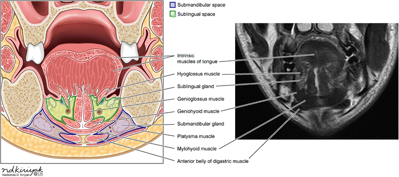
Identify the mylohyoid and compare the sublingual and submandibular compartments in the schematic and normal MRI. Relate the compartments to the tooth-apex relationships discussed in this section.
Coronal sublingual and submandibular spaces with normal MRI correlation · Swapnil Patel and Alok A. Bhatt (2018); illustration credited within the figure to Nadezhda D. Kiriyak / University of Rochester · CC BY 4.0. Unchanged published image; displayed responsively. Original publication.
Assess severity before choosing a dental intervention
Record observations when appropriate, including temperature, pulse, respiratory rate, blood pressure, oxygen saturation and mental state. Ask about speed of progression, swallowing, breathing, voice, oral intake and urine output. Examine mouth opening, floor of mouth, neck, facial spread and the eye region where relevant.
Rapid progression, significant trismus, floor-of-mouth swelling, dysphagia, breathing difficulty or deep/high-risk space involvement require urgent hospital assessment. Immunocompromise and reduced physiological reserve lower the threshold for concern. Absence of fever cannot exclude serious infection, especially in an older or immunosuppressed person.
Recognise sepsis without reducing it to a temperature
Sepsis is life-threatening organ dysfunction caused by a dysregulated response to infection. Confusion, poor perfusion, respiratory compromise and reduced urine output can be more important than the size of the facial swelling. Apply the correct sepsis pathway to the patient and clinical setting.
NICE NG253 concerns people aged 16 or over who are not pregnant or recently pregnant; separate guidance applies to children and pregnancy. NEWS2 has a role in appropriate acute settings but does not replace clinical judgement or detect every anatomical airway threat. A threatened airway must not wait for CT or a laboratory result.
Achieve source control
For a suitable stable patient, drainage may be achieved through the tooth, extraction or incision of an accessible collection. Choose the route according to diagnosis, restorability, anatomy and competence. A collection does not resolve reliably if its cause remains untreated, and repeated prescriptions can allow disease to progress.
Patients with extensive infection may need coordinated surgical drainage and airway management in hospital. Blood tests and contrast-enhanced imaging can help assess systemic impact, collection and extent when clinically indicated. Obtain investigations according to stability; do not allow a desired scan to delay resuscitation or emergency transfer.
Prescribe antibiotics for an indication and review response
SDCEP prioritises local measures. Antibiotics are indicated for spreading infection, systemic involvement or situations where immediate drainage cannot be achieved according to the current pathway. Choose the medicine using current guidance, allergy assessment, interactions, organ function and relevant microbiological advice; broadening therapy is not a substitute for reassessment.
Record the indication and arrange review, ideally at three days for patients prescribed antibiotics in the SDCEP dental abscess pathway. At review, assess systemic signs and source control and follow its response-based stopping advice. Deterioration needs earlier contact and may change the care setting; do not ask the patient to wait for a scheduled review.
Prevent recurrence and communicate the safety net
Explain the cause, what has been drained or removed and any remaining definitive treatment. Check analgesia, hydration and ability to obtain care. Give explicit warning signs and emergency contacts. Where emergency transfer is needed, communicate observations, airway concerns, progression, comorbidity, allergies and medicines already given.
After acute recovery, complete the restorative, endodontic or extraction plan and address prevention. A quieter tooth after antibiotics is not evidence that the source has healed. Persistent or recurrent swelling despite treatment should reopen the differential and prompt appropriate specialist assessment, including non-odontogenic pathology.
A distinction to remember
A normal temperature and a small visible swelling do not rule out serious disease. Antibiotics cannot compensate for an unprotected airway or an untreated source.
A patient has a local fluctuant swelling from a non-restorable tooth but is otherwise well. What is the central treatment?
Reveal the reasoning
Provide appropriate source control and drainage, including extraction when indicated and within competence, with suitable analgesia. Antibiotics are not routinely needed for a localised infection adequately managed by local measures. Assess the patient for spread and give a clear deterioration safety net.
Test your recall
What determines the initial direction of odontogenic infection spread?
Show answer
The site of cortical perforation and its relationship to muscle attachments and fascial spaces influence spread. For lingual mandibular infection, the mylohyoid relationship helps explain sublingual versus submandibular involvement. Actual examination remains essential because infection can extend beyond its first compartment.