
Jaw cysts and lesions
A radiolucency is a radiographic description, not a diagnosis of a cyst. The tooth relationship, internal pattern, behaviour and clinical findings narrow the differential. Management then follows the lesion’s biology, anatomical extent and histology, with follow-up long enough to detect recurrence or altered behaviour.
Quick revision summary
Classify the process before naming a lesion
A true cyst has a pathological cavity with an epithelial lining; some jaw cavities are pseudocysts without that lining. Odontogenic cysts arise from tooth-related epithelial remnants and can be inflammatory or developmental.
Choose investigations for extent and diagnosis
An intraoral or panoramic image may show a lesion but not its full buccolingual extent. CBCT or other cross-sectional imaging can answer specific bony or soft-tissue questions. Select imaging according to anticipated management, avoiding routine additional exposure that cannot change care.
Review the biological outcome
Record the final diagnosis, operation, specimen details and any residual concern. Follow-up examines symptoms, clinical expansion, tooth effects and radiographic healing, with intervals and duration matched to lesion and treatment.
Common mistake
Vitality and pericoronal relationships narrow a differential; neither can prove the histological identity of a jaw lesion.
Jaw cysts and lesions: construct and test a differential
A radiolucency is a radiographic description, not a diagnosis of a cyst. The tooth relationship, internal pattern, behaviour and clinical findings narrow the differential. Management then follows the lesion’s biology, anatomical extent and histology, with follow-up long enough to detect recurrence or altered behaviour.
Classify the process before naming a lesion
A true cyst has a pathological cavity with an epithelial lining; some jaw cavities are pseudocysts without that lining. Odontogenic cysts arise from tooth-related epithelial remnants and can be inflammatory or developmental. Jaw tumours, giant-cell lesions, fibro-osseous disease, infection and malignancy can mimic cystic appearances.
Use the clinical setting to rank alternatives rather than memorising a single image-to-diagnosis match. A small periapical radiolucency may fit inflammatory disease, while an expanding multilocular lesion requires a broader assessment. Well-defined borders often suggest slower growth, but do not establish that the lesion is harmless.
Tooth & supporting tissues
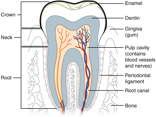
Trace the pulp and root canal to the apical region, then identify the periodontal ligament and supporting bone. Relate these structures to avulsion, apical pathology and extraction. The original uses the spelling “dentin”; cementum is not separately labelled.
Tooth & supporting tissues · OpenStax College · CC BY 3.0. Original image; displayed at a responsive size. Original publication.
Use tooth relationships accurately
A radicular cyst is associated with a non-vital tooth in the appropriate inflammatory context, while a residual cyst remains after the causative tooth is removed. A dentigerous cyst relates to the crown of an unerupted tooth and attaches around the cervical region. An eruption cyst is a soft-tissue counterpart over an erupting tooth.
An odontogenic keratocyst may be pericoronal or unrelated to an impacted crown and can extend through the jaw with relatively limited expansion. A pericoronal lesion could also represent ameloblastoma or another entity. Tooth position is an anatomical clue, not proof of the lining’s identity.
Dentigerous cyst: a pericoronal radiolucency
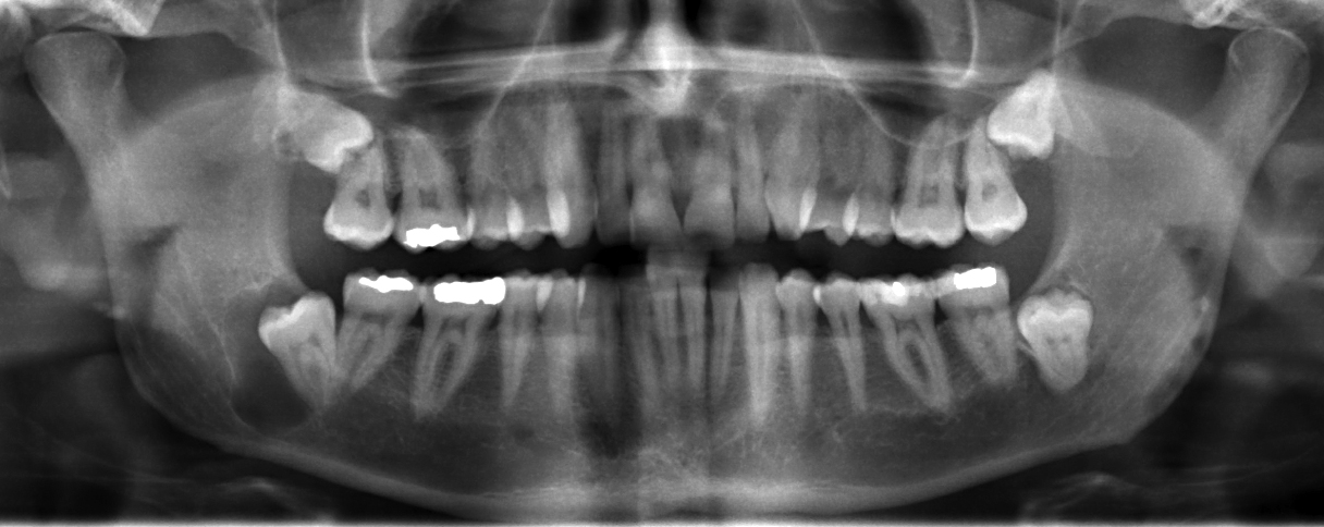
Locate the radiolucency associated with the impacted lower third molar on the left of the displayed image; the source describes a right mandibular dentigerous cyst. Assess its border and relationship to the crown, adjacent roots and mandibular canal. A pericoronal radiolucency supports a differential; imaging alone cannot establish the lining or exclude other odontogenic lesions.
JawCyst.jpg · Coronation Dental Specialty Group · CC BY 3.0. Source image reproduced without alteration; any arrows or circles are part of the original.
Which tooth relationship makes a dentigerous cyst an important consideration?
A radiolucency surrounding the crown of an unerupted tooth. An enlarged follicle and other odontogenic lesions can resemble it, so clinical, radiographic and appropriate histological findings must be correlated.
Describe the lesion reproducibly
Record jaw and side, anteroposterior extent, relation to teeth, number, border and transition zone. Describe unilocular or multilocular architecture, internal radiopacity or septa and effects on surrounding structures. Root displacement, resorption, canal displacement, expansion and cortical destruction help assess behaviour.
Examine swelling, mucosal change, tenderness, drainage, tooth vitality and mobility, occlusion and sensation. Ask about growth, pain, previous lesions and systemic symptoms. Unexplained numbness, rapid progression, ill-defined destruction or a soft-tissue mass increases concern and should influence urgency and investigation.
Odontogenic keratocyst: a posterior mandibular example
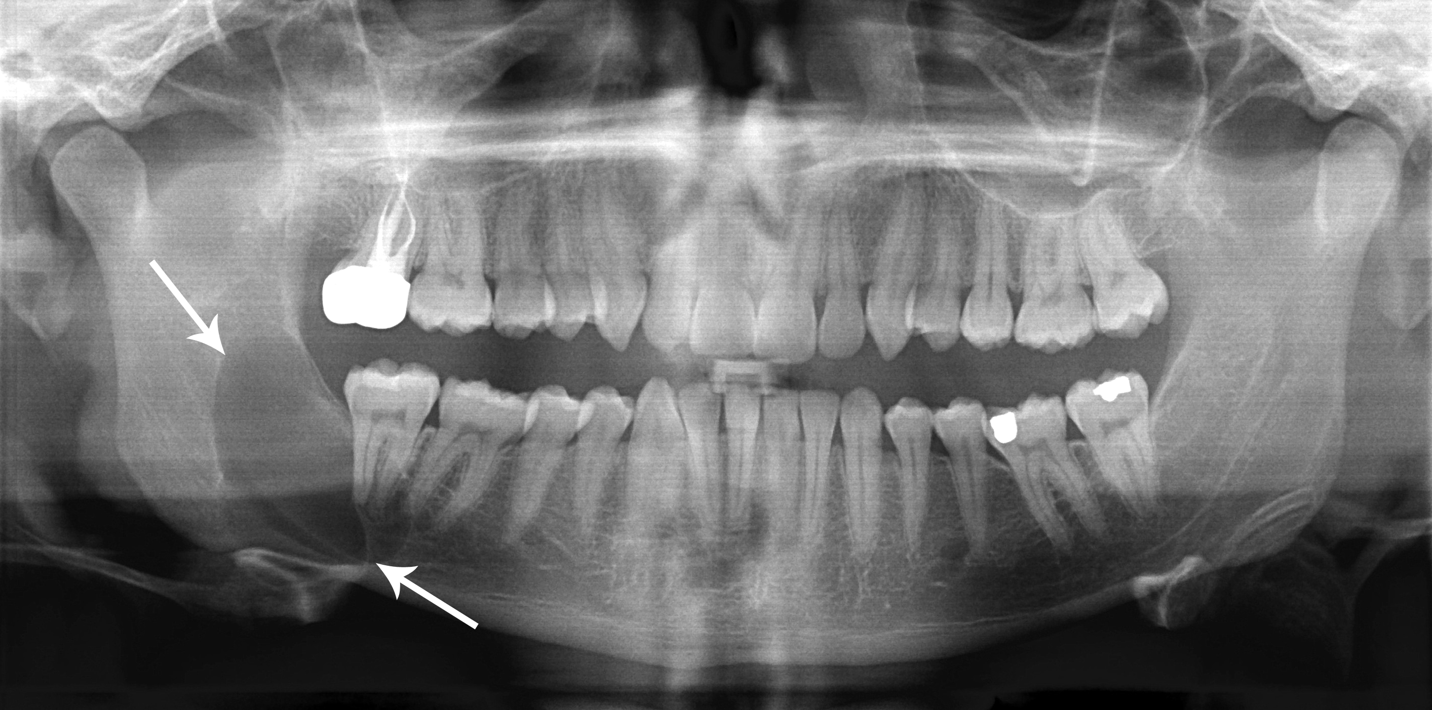
Use the original arrows to locate the posterior mandibular radiolucency and inspect its extent. The source diagnosis is an odontogenic keratocyst, described under the historical name “keratocystic odontogenic tumour”. Its relatively well-defined appearance does not make follow-up unnecessary. Other jaw lesions can look similar; definitive classification requires correlation with tissue findings.
Classic_keratocystic_odontogenic_tumour.jpg · Coronation Dental Specialty Group · CC BY 3.0. Source image reproduced without alteration; any arrows or circles are part of the original.
Can the relatively well-defined border distinguish this keratocyst from every other odontogenic cyst?
No. Borders and location help rank possibilities but overlap between lesions. The source diagnosis is supplied for teaching; it cannot be independently established from this panoramic image alone.
Choose investigations for extent and diagnosis
An intraoral or panoramic image may show a lesion but not its full buccolingual extent. CBCT or other cross-sectional imaging can answer specific bony or soft-tissue questions. Select imaging according to anticipated management, avoiding routine additional exposure that cannot change care.
Histology and clinical correlation establish most definitive lesion diagnoses. Aspiration may support assessment or alert to vascularity, but fluid appearance alone is not diagnostic. When vascular pathology is plausible, obtain appropriate specialist assessment before invasive sampling. A small biopsy can miss heterogeneous tumour components, so discordant findings need review.
Nasopalatine duct cyst: an anterior maxillary example
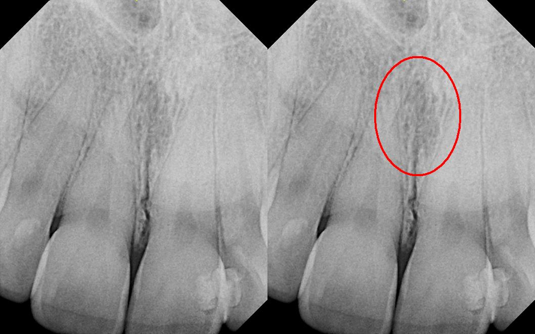
The right panel repeats the image with the source’s red circle highlighting the midline region. The source explicitly corrects its filename: this is reported as a nasopalatine duct cyst, not a nasolabial cyst. Consider the incisive canal and adjacent incisors, including pulp tests. The subtle appearance illustrates why a labelled example is not a stand-alone diagnostic rule.
Nasolabial_duct_cyst.JPG · DRosenbach · CC BY-SA 3.0. Source image reproduced without alteration; any arrows or circles are part of the original.
Why assess the adjacent incisors before treating a midline anterior maxillary radiolucency as endodontic disease?
A nasopalatine duct cyst or normal incisive-canal anatomy may overlap the incisor roots. Clinical examination, pulp tests and appropriate imaging help avoid unnecessary endodontic treatment of otherwise healthy teeth.
Compare enucleation, decompression and resection
Enucleation removes the cystic lining where feasible. Decompression or marsupialisation reduces pressure and can permit bone formation or reduce morbidity near important structures, but requires adherence, hygiene and prolonged review. Reduction in size does not prove that all pathological tissue has disappeared.
Large size alone does not determine treatment: pathology, recurrence risk, proximity to nerves or teeth, fracture risk and the patient’s circumstances matter. Some lesions require adjunctive treatment or resection because of biological behaviour. These choices belong to a specialist plan balancing disease control with functional and anatomical morbidity.
Ameloblastoma: expansion on axial CT
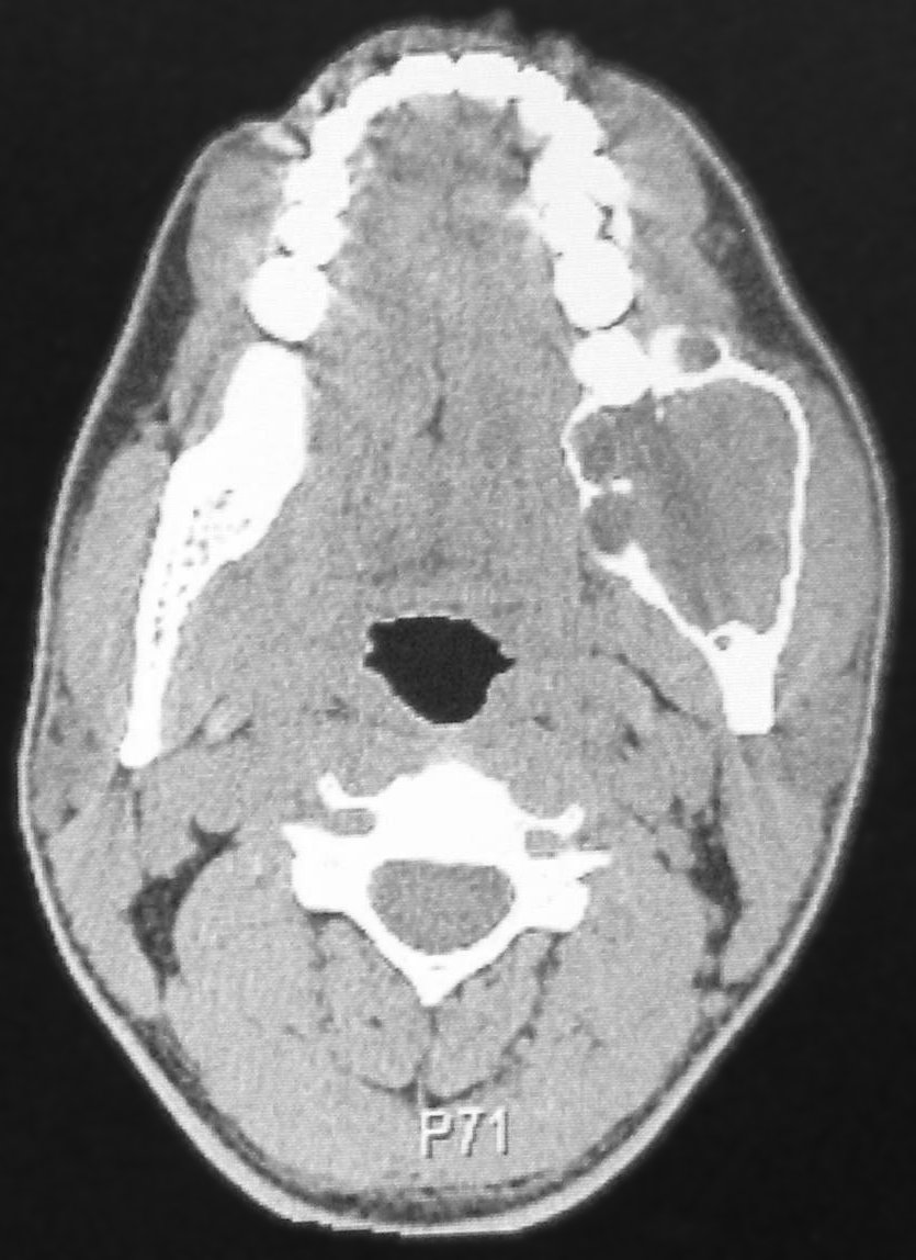
Compare the expanded posterior mandible on the right of the displayed image with the opposite side. The source reports a left mandibular ameloblastoma. Trace the remaining cortex and internal compartmental appearance. This CT example and the histology example elsewhere in the atlas come from separate teaching cases.
CT Scan of ameloblastoma.jpg · Berto1286; source-file crop and tonal adjustment by Hellerhoff · Public domain. Reproduced without alteration. Labels, arrows and circles, where present, belong to the source image.
What does this axial view show that a panoramic projection may conceal?
Buccolingual expansion and the relationship to the cortical boundaries are more directly visible. The imaging pattern informs the differential and extent assessment; tissue findings establish classification.
Understand keratocyst recurrence and syndromic clues
Odontogenic keratocysts have a recurrence tendency related to their lining, possible satellite cysts and treatment factors. Treatment may include selected adjuncts through specialist services, with their own risks. Do not present one recurrence percentage or one adjunct as universally applicable across techniques and populations.
Multiple keratocysts or a young presentation can prompt assessment for Gorlin syndrome, alongside features such as basal cell carcinomas, palmar pits and relevant family history. Not every keratocyst is syndromic. Appropriate genomic assessment and counselling should follow clinical criteria rather than an isolated dental assumption.
Stafne bone cavity: panoramic location
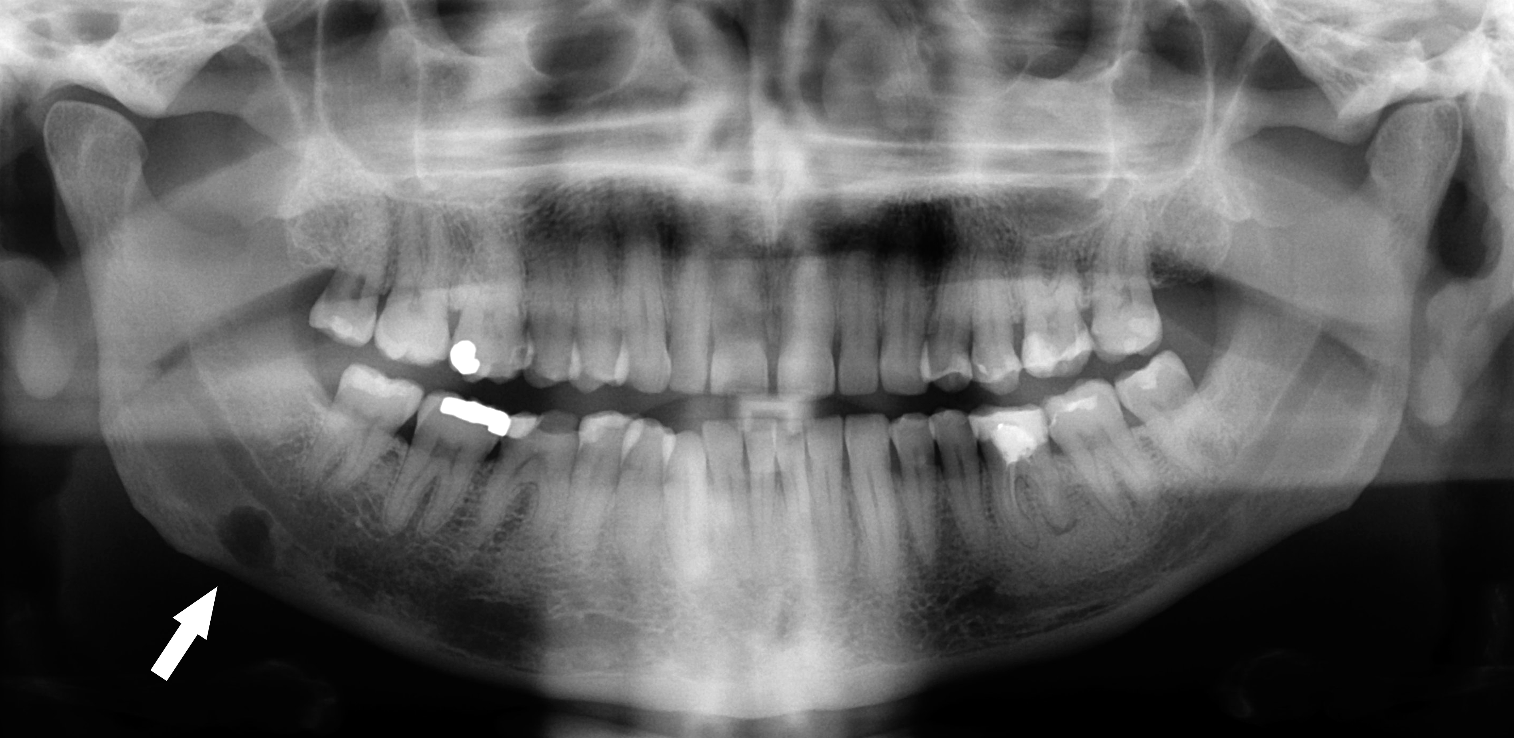
Follow the original arrow to the rounded radiolucency near the lower border of the right mandible, on the left of the displayed image. The source identifies a Stafne defect. Compare its position below the mandibular canal with the tooth-related relationships of odontogenic cysts. The axial CT shows the same source case from another plane.
Stafne defect panorex.jpg · Coronation Dental Specialty Group · CC BY 3.0. Reproduced without alteration. Labels, arrows and circles, where present, belong to the source image.
Which anatomical relationship is particularly helpful in this example?
Its location below the mandibular canal near the posterior lower border. Use the full pattern and clinical context; that relationship alone is not a universal rule for every radiolucency.
Review the biological outcome
Record the final diagnosis, operation, specimen details and any residual concern. Follow-up examines symptoms, clinical expansion, tooth effects and radiographic healing, with intervals and duration matched to lesion and treatment. Long-term recurrence surveillance may be needed even after mucosal healing.
A new or enlarging area, recurrent discharge or sensory change should prompt reassessment. Explain the expected course and why attendance matters, especially during decompression. If the histology does not fit the imaging or clinical behaviour, seek specialist radiology and pathology review instead of assuming that the original label remains correct.
Stafne bone cavity: the lingual depression on CT
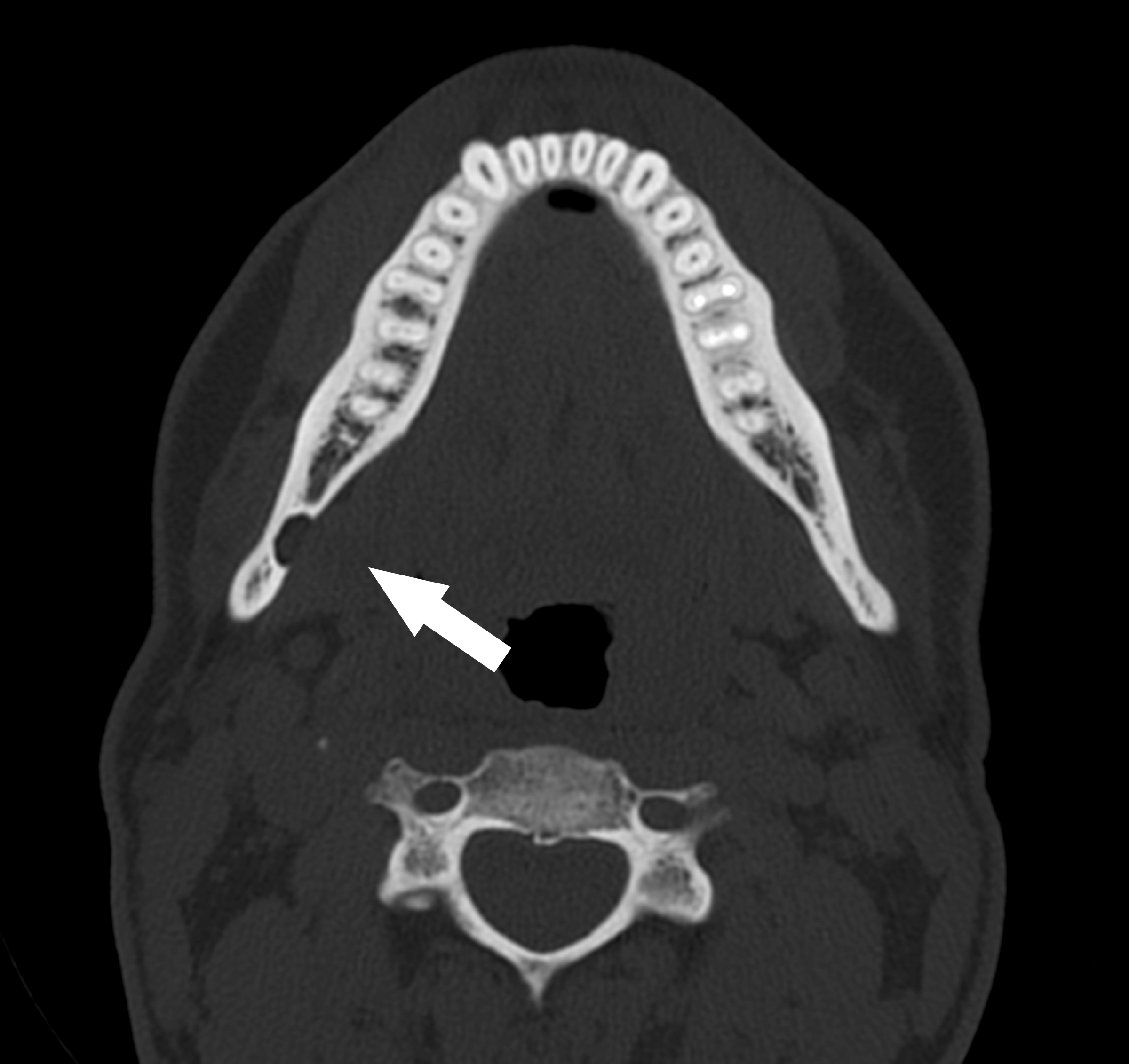
The original arrow marks a concavity in the lingual mandibular cortex. Compare it with the panoramic example of the same source case. This helps explain why a two-dimensional radiolucency can represent a bony depression rather than an epithelial-lined odontogenic cyst. Further imaging in a real case needs an individual clinical indication.
Stafne defect CT axial.jpg · Coronation Dental Specialty Group · CC BY 3.0. Reproduced without alteration. Labels, arrows and circles, where present, belong to the source image.
Why should a Stafne bone cavity not automatically be called a true odontogenic cyst?
It represents a mandibular bony depression rather than an epithelial-lined tooth-derived cyst. Classification depends on the complete assessment.
Cemento-osseous dysplasia: localised and florid examples
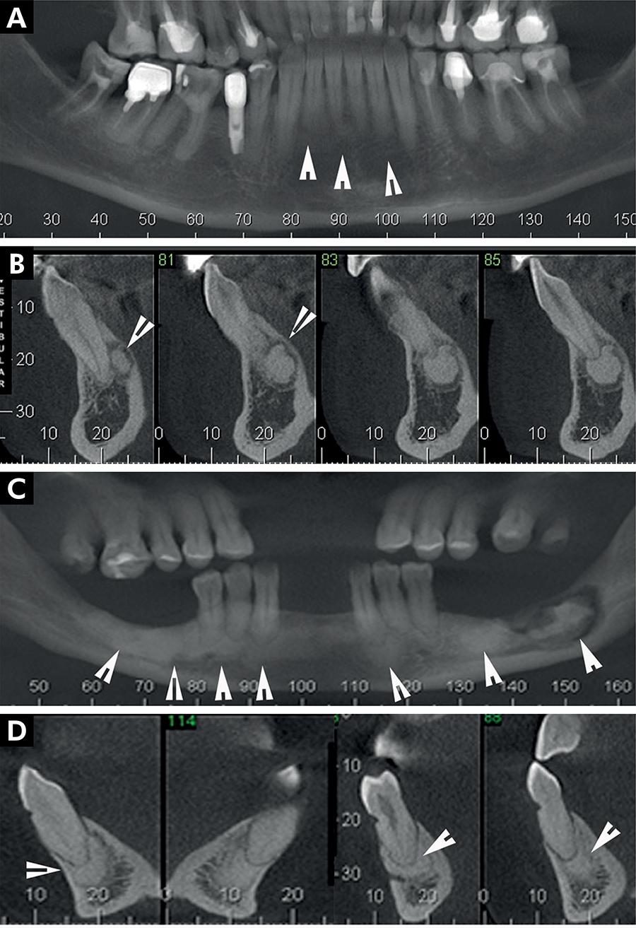
Compare the distribution and separation from roots in the source examples.
Panel key
- A–B
- Periapical cemento-osseous dysplasia.
- C–D
- Florid cemento-osseous dysplasia; the source also describes sequestration in C.
Periapical radiopaque-hyperdense jaw lesions 2.jpg · Brunno Santos Freitas Silva, Mike Reis Bueno, Fernanda P. Yamamoto-Silva, Ricardo Santiago Gomez, Ove Andreas Peters and Carlos Estrela · CC BY 4.0. Reproduced without alteration. Labels, arrows and circles, where present, belong to the source image. Original publication.
Why assess the teeth before attributing these findings to pulp disease?
The imaging distribution may have a non-endodontic explanation.
Fibrous dysplasia, osteoma and cementoblastoma
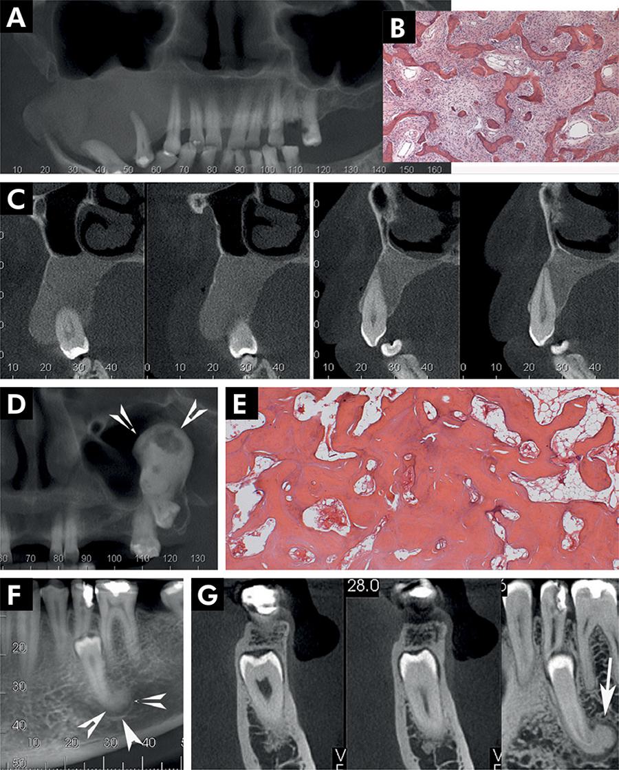
Compare the margins and root relationships of these source cases.
Panel key
- A–C
- Fibrous dysplasia.
- D–E
- Central osteoma.
- F–G
- Cementoblastoma.
Periapical radiopaque-hyperdense jaw lesions 3.jpg · Brunno Santos Freitas Silva, Mike Reis Bueno, Fernanda P. Yamamoto-Silva, Ricardo Santiago Gomez, Ove Andreas Peters and Carlos Estrela · CC BY 4.0. Reproduced without alteration. Labels, arrows and circles, where present, belong to the source image. Original publication.
Which relationship deserves attention in the cementoblastoma panels?
The lesion’s attachment to the root.
Odontomas and other calcified jaw lesions
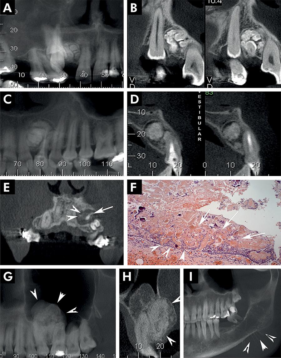
Use the panel key to compare the source diagnoses. The historical label CCOT corresponds here to calcifying odontogenic cyst.
Panel key
- A–B
- Compound odontoma.
- C–D
- Complex odontoma.
- E–F
- Calcifying odontogenic cyst (source label: CCOT).
- G–H
- Adenomatoid odontogenic tumour.
- I
- Source example of osteosarcoma.
Periapical radiopaque-hyperdense jaw lesions 4.jpg · Brunno Santos Freitas Silva, Mike Reis Bueno, Fernanda P. Yamamoto-Silva, Ricardo Santiago Gomez, Ove Andreas Peters and Carlos Estrela · CC BY 4.0. Reproduced without alteration. Labels, arrows and circles, where present, belong to the source image. Original publication.
Does calcification establish benignity?
No. Compare the entire lesion and clinical findings.
Ameloblastoma: epithelial islands on histology
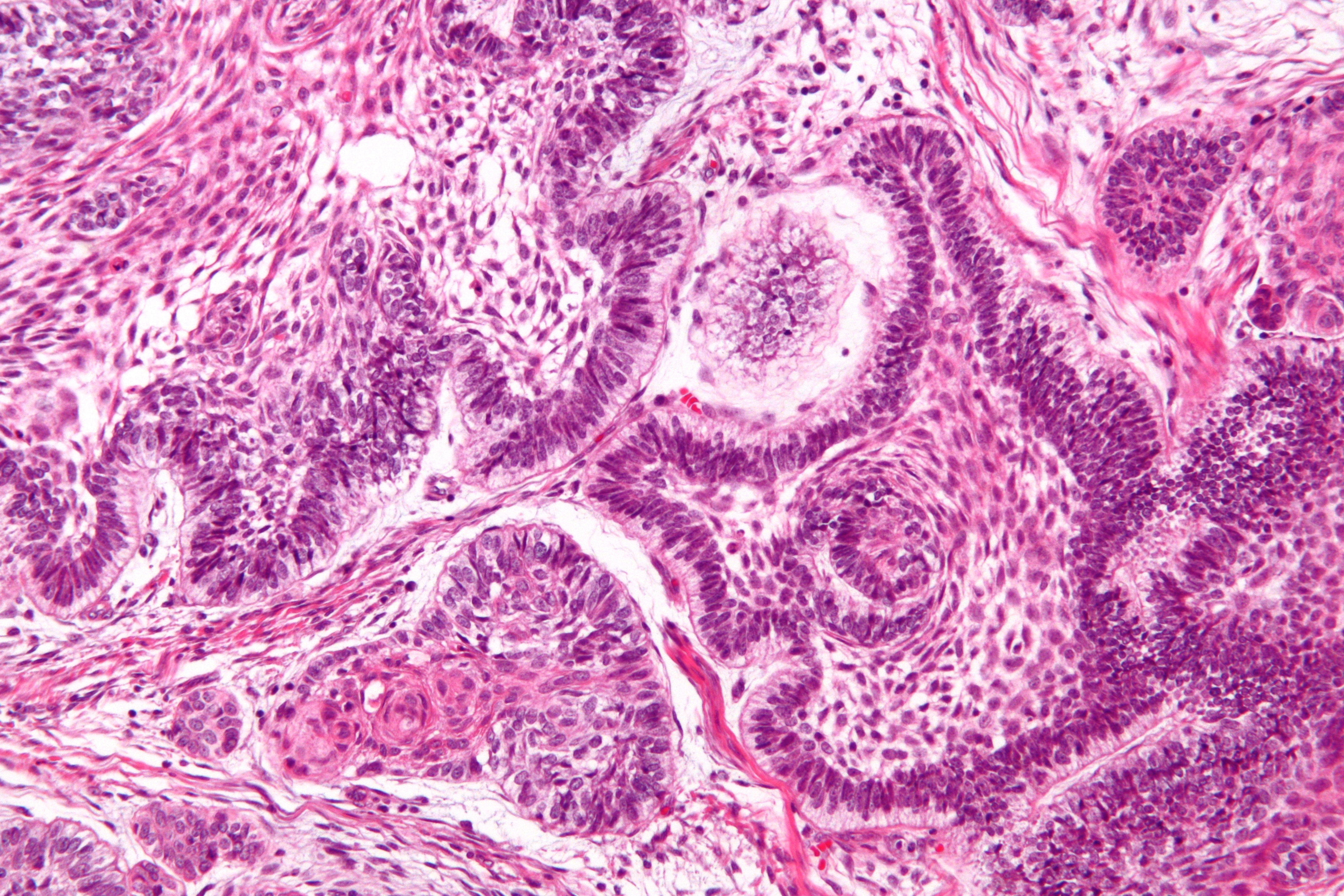
Locate the epithelial islands within the connective-tissue stroma. The source identifies peripheral palisading, reverse nuclear polarisation and central stellate-reticulum-like cells. This is a separate specimen from the CT teaching case. A complete diagnosis considers the sampled architecture and the clinical and radiographic findings.
Ameloblastoma - high mag.jpg · Nephron · CC BY-SA 3.0. Reproduced without alteration. Labels, arrows and circles, where present, belong to the source image.
What does reverse nuclear polarisation refer to in this teaching example?
The nuclei of peripheral cells lie away from the basement-membrane side. It is interpreted with the wider architecture rather than treated as an isolated diagnostic rule.
Odontoma: dental hard-tissue architecture
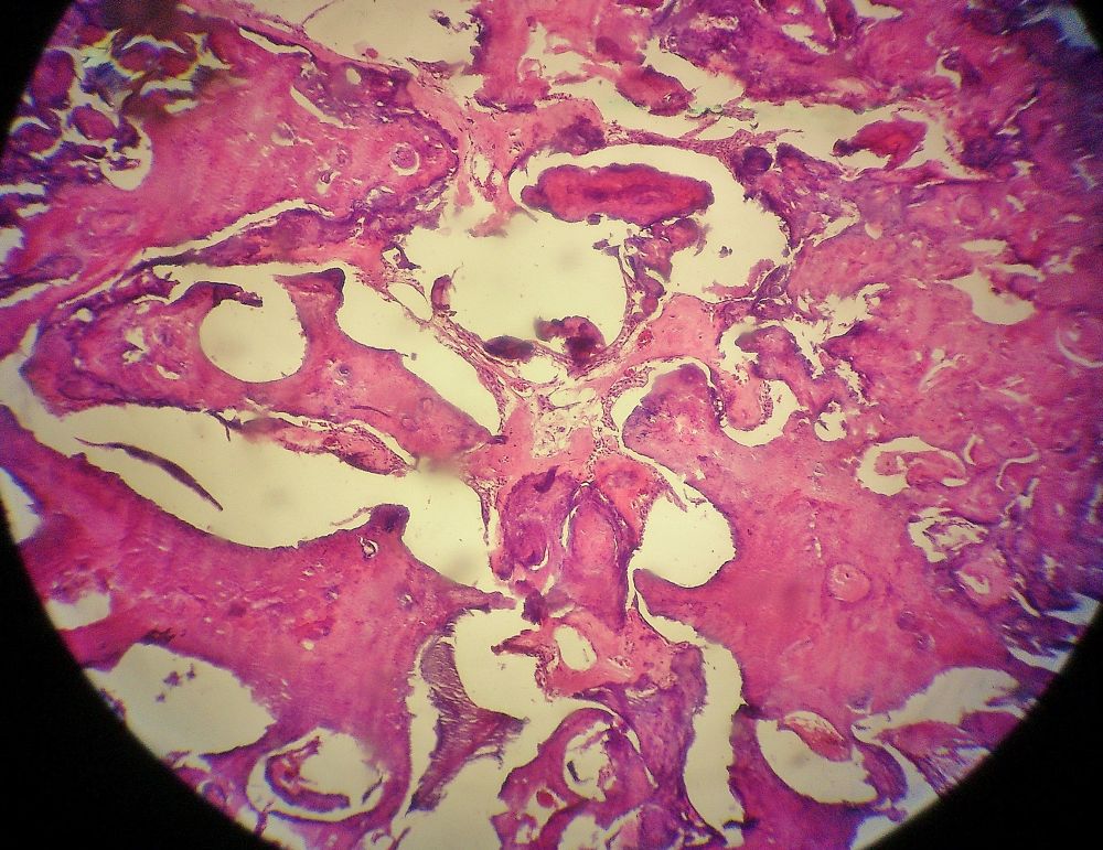
The source identifies an odontoma. Inspect how the dental tissues are organised and compare this tissue view with the atlas’s radiographic odontoma panels, which show different teaching cases. Imaging describes the mineralised pattern and tooth relationships; histology assesses the tissue architecture.
Odontoma.jpg · JXtra · CC BY-SA 3.0. Reproduced without alteration. Labels, arrows and circles, where present, belong to the source image.
Why keep the radiographic and histological descriptions distinct?
They describe different aspects of the lesion. A radiopaque pattern is not itself a microscopic diagnosis, and the specimen must be interpreted with the case information.
Radicular cyst: epithelial metaplasia
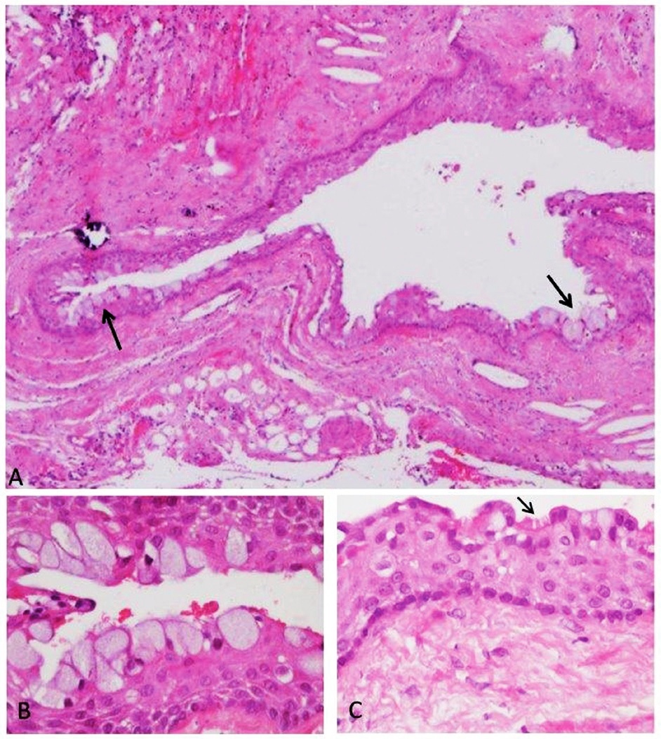
The source demonstrates metaplastic epithelial changes in a radicular-cyst wall. These are selected examples, not a description of every radicular cyst. Interpret unusual lining features with the rest of the architecture and the clinical relationship to the teeth.
Panel key
- A
- Cyst wall with mucous-cell changes.
- B
- Higher-power view of mucous cells.
- C
- Ciliated epithelial cells, marked by the source arrow.
Histopathology of a periapical cyst, with metaplastic changes of mucous secreting cells, and ciliated cells.jpg · Igor Tsesis, Eyal Rosen, Liz Dubinsky, Amos Buchner and Marilena Vered · CC BY 2.5. Reproduced without alteration. Labels, arrows and circles, where present, belong to the source image. Original publication.
Do mucous cells alone establish a glandular odontogenic cyst?
No. Mucous-cell metaplasia can occur in other cysts. Classification requires the complete histological pattern and clinical correlation.
A distinction to remember
Vitality and pericoronal relationships narrow a differential; neither can prove the histological identity of a jaw lesion.
A well-defined radiolucency lies beside the apex of a vital tooth. Should it automatically receive root canal treatment?
Reveal the reasoning
No. Confirm the sensibility findings and consider anatomical structures and non-endodontic lesions. A neighbouring lesion is not necessarily caused by that tooth. Use the clinical pattern and appropriate imaging or referral before undertaking irreversible treatment without a supported pulpal diagnosis.
Test your recall
How do radicular and dentigerous cysts differ in tooth relationship?
Show answer
A radicular cyst arises in an inflammatory context around a non-vital tooth; a dentigerous cyst surrounds the crown of an unerupted tooth around its cervical attachment. These relationships guide the differential, but clinical assessment and appropriate pathological confirmation remain necessary.