
Screening and full charting
Screening identifies who needs investigation; detailed charting establishes the distribution and severity of disease and allows response to be measured. A useful record links the measurements to diagnosis, treatment and a defined review plan.
Quick revision summary
Screen, then investigate
BPE records the highest sextant finding. Code 3 leads to affected-sextant charting after initial treatment; code 4 requires whole-mouth charting before treatment. Furcation findings also need fuller assessment.
Measure the right things
Use six sites, gentle technique and a stable attachment reference. Pocket depth changes with gingival-margin position, so a shallower pocket alone does not demonstrate regeneration or erase previous loss.
Monitor established disease
Periodontitis needs site-level review, including a full-mouth assessment at least annually. Link change to plaque, inflammation, risk and anatomy, then document the treatment or review decision.
Common mistake
A BPE score is neither a diagnosis nor a maintenance chart. It cannot demonstrate attachment gain or locate the surface needing treatment.
Screening and full charting
Screening identifies who needs investigation; detailed charting establishes the distribution and severity of disease and allows response to be measured. A useful record links the measurements to diagnosis, treatment and a defined review plan.
Use BPE as a screen
Examine adult sextants with a ball-ended WHO probe and light force, walking around all relevant surfaces. The black band extends from 3.5 to 5.5 mm. Record the highest code per sextant, with an asterisk for furcation involvement. A sextant normally requires at least two teeth; include a lone tooth with its neighbour. Third molars are generally excluded unless replacing absent first or second molars.
Code 0 indicates no bleeding, calculus or pocketing above the screen threshold; 1 indicates bleeding; 2 calculus or plaque-retentive margins with the band visible; 3 a partly obscured band; and 4 a completely obscured band. Continue examining the sextant after finding code 4 to avoid missing other findings. Age-appropriate protocols apply to children and young people rather than simply using the adult pathway.
Periodontitis: pocket formation and loss of support
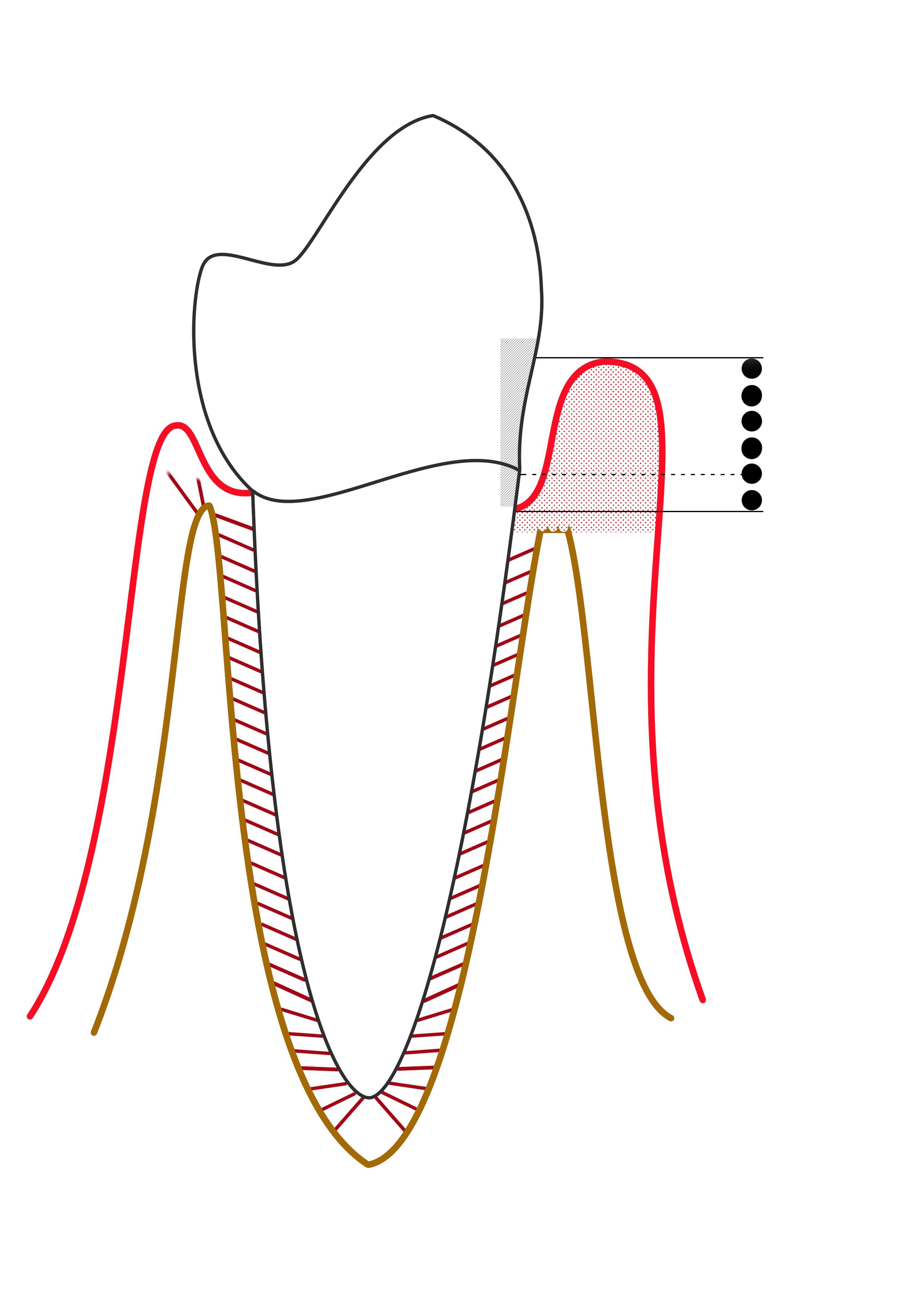
Compare the apical extent of the pocket and the reduced supporting bone on the affected side. This simplified diagram is labelled “mild periodontitis” by its author; it is not a modern stage/grade classification chart.
Mild periodontitis.png · Ian Furst · CC BY-SA 4.0. None; reproduced without alteration.
Why is probing depth different from clinical attachment level?
Probing depth runs from the gingival margin to the pocket base. Clinical attachment level uses the cemento-enamel junction as its reference; recession or enlargement changes their relationship.
Know when screening is insufficient
A sextant code cannot identify the exact tooth, surface, depth or attachment level. It also cannot distinguish swollen gingiva from destructive disease. Current SDCEP guidance recommends detailed charting of a BPE 3 sextant after initial treatment; BPE 4 requires whole-mouth charting before treatment. Furcation involvement also prompts a fuller examination.
An established periodontitis patient needs detailed review rather than repeated BPE screening alone. A sequence of improving box charts cannot demonstrate site-level stability or attachment gain. Document the disease history even when treatment has reduced the screening codes, and explain why more detailed monitoring remains relevant.
Measure six sites and a stable reference
Record mesiobuccal, midbuccal, distobuccal, mesiolingual, midlingual and distolingual findings. Follow the root with gentle walking strokes, angling interproximally to reach beneath the contact. Probe force, diameter, angulation, tissue inflammation and deposits all affect readings. Forcing the probe or sampling only the midbuccal surface produces misleading information.
Probing depth is measured from the gingival margin; attachment level uses a stable reference such as the CEJ. Recession or a coronal gingival margin changes their relationship. A smaller pocket after treatment may reflect tissue shrinkage rather than regeneration. Record enough margin information to interpret the change instead of assuming every numerical reduction represents attachment gain.
Record features that change prognosis
Assess furcation entrances with an appropriate curved probe and describe horizontal involvement. Add mobility, suppuration, recession, migration, plaque and missing teeth. Examine cleaning access and relevant restoration margins. Radiographs may suggest furcation loss but superimposition can conceal or exaggerate the apparent defect; combine them with clinical findings.
An isolated narrow deep pocket at a root-filled tooth deserves a focused differential, including fracture or endodontic drainage. Generalised broad pocketing has a different context. Mobility can reflect inflammation, reduced support or loading, so it should not be translated automatically into a hopeless prognosis or an instruction to adjust the occlusion.
Make review comparable with baseline
Use a consistent sequence, legible site labels and a comparable probing technique. SDCEP review charting assesses pocket depth and bleeding at six sites per tooth, records depths of at least 4 mm and bleeding sites, and requires a full-mouth assessment at least annually in periodontitis. Add other features when they help interpret change.
A small isolated difference can be measurement variation. Reproducible depth increases, suppuration, attachment change and relevant radiographic trends strengthen concern. The measurement should lead to an explanation and response: additional cleaning support, focused treatment, further investigation or referral. A chart without a diagnostic interpretation is an incomplete clinical outcome.
Select images for the remaining question
A horizontal bitewing may exclude the crest in a patient with recession or advanced loss. Depending on distribution, periapicals, vertical bitewings or an appropriate panoramic examination may better show support and root anatomy. Existing suitable images can sometimes answer the question without another exposure.
For example, several bleeding 6 mm molar pockets need site-level assessment and a treatment plan, not just a BPE 4 entry. A single atypical defect needs an explanation before it determines whole-mouth classification. Record the diagnosis, shared decisions and review endpoint alongside the chart so the next clinician can understand the reasoning.
Periodontal bone loss on a periapical radiograph
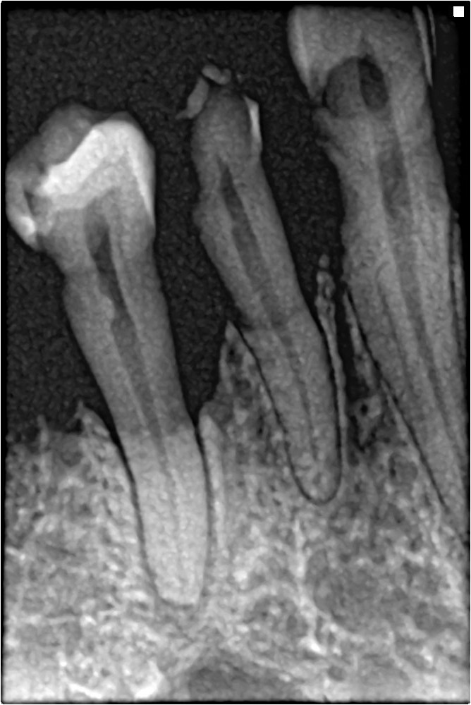
Trace the reduced bone height and irregular contour between the teeth. Correlate with full periodontal charting; this single image cannot establish the patient’s complete stage and grade.
Bone loss in periapical xray.jpg · Shaimaa Abdellatif · CC BY-SA 4.0. Image reproduced without alteration; original marks and any embedded credits retained.
A distinction to remember
A BPE score is neither a diagnosis nor a maintenance chart. It cannot demonstrate attachment gain or locate the surface needing treatment.
One sextant scores BPE 3 and none score 4. When is detailed charting done under SDCEP advice?
Reveal the reasoning
Chart the affected sextant after initial treatment. The initial assessment still needs to identify disease and other care needs. If any sextant scores 4, whole-mouth detailed charting is required before treatment begins rather than following the same code-3 timing.
Test your recall
What does a BPE 4 tell you, and what does it leave unknown?
Show answer
It identifies a screening depth beyond the visible black band, but not the exact tooth, surface, depth or attachment level. It also compresses all other sites into one score. Detailed charting is needed to locate disease and plan treatment.