
Periodontal health and gingivitis
Distinguish reversible gingival inflammation from destruction of the attachment apparatus. A useful assessment separates today’s inflammation, historical support loss and the factors that will determine whether control can be maintained.
THE ANATOMY LAB / INTERACTIVE 3DStudy the tooth’s supporting tissues in 3D↗Quick revision summary
Separate inflammation from destruction
Gingivitis can resolve with effective plaque control. Periodontitis leaves attachment loss even when inflammation settles. Measure pockets, recession and bleeding together rather than deciding from gingival colour or one depth.
Check the context
Smoking can conceal bleeding; hormones and medicines can amplify gingival changes. Unusual ulceration, peeling or disproportionate bleeding requires a broader diagnosis rather than another routine cleaning appointment.
Make daily care achievable
Demonstrate aids at the sites that need them, remove deposits and correct access barriers. Review the tissue response and retain the historical diagnosis when the periodontium is reduced.
Common mistake
Bleeding describes current inflammation; it does not establish how much periodontal support has already been lost.
Periodontal health and gingivitis
Distinguish reversible gingival inflammation from destruction of the attachment apparatus. A useful assessment separates today’s inflammation, historical support loss and the factors that will determine whether control can be maintained.
How plaque produces gingivitis
The acquired pellicle forms on a cleaned tooth and permits bacterial attachment. A maturing biofilm is an organised community rather than a collection of isolated bacteria. Microbial products stimulate the gingival host response: vessels dilate, inflammatory cells accumulate and the sulcular epithelium becomes more vulnerable to bleeding. Calculus is mineralised plaque and a plaque-retentive surface, not a replacement explanation for the living biofilm around it.
Clinical changes include redness, rounded papillae, loss of stippling, swelling and bleeding with gentle probing. Swelling can deepen the sulcus without loss of attachment, producing a gingival or false pocket. The expression varies with smoking, hormones, medicines and susceptibility. Little visible plaque on the examination day does not reconstruct the patient’s previous exposure.
Gingivitis: inflamed gingiva without attachment loss
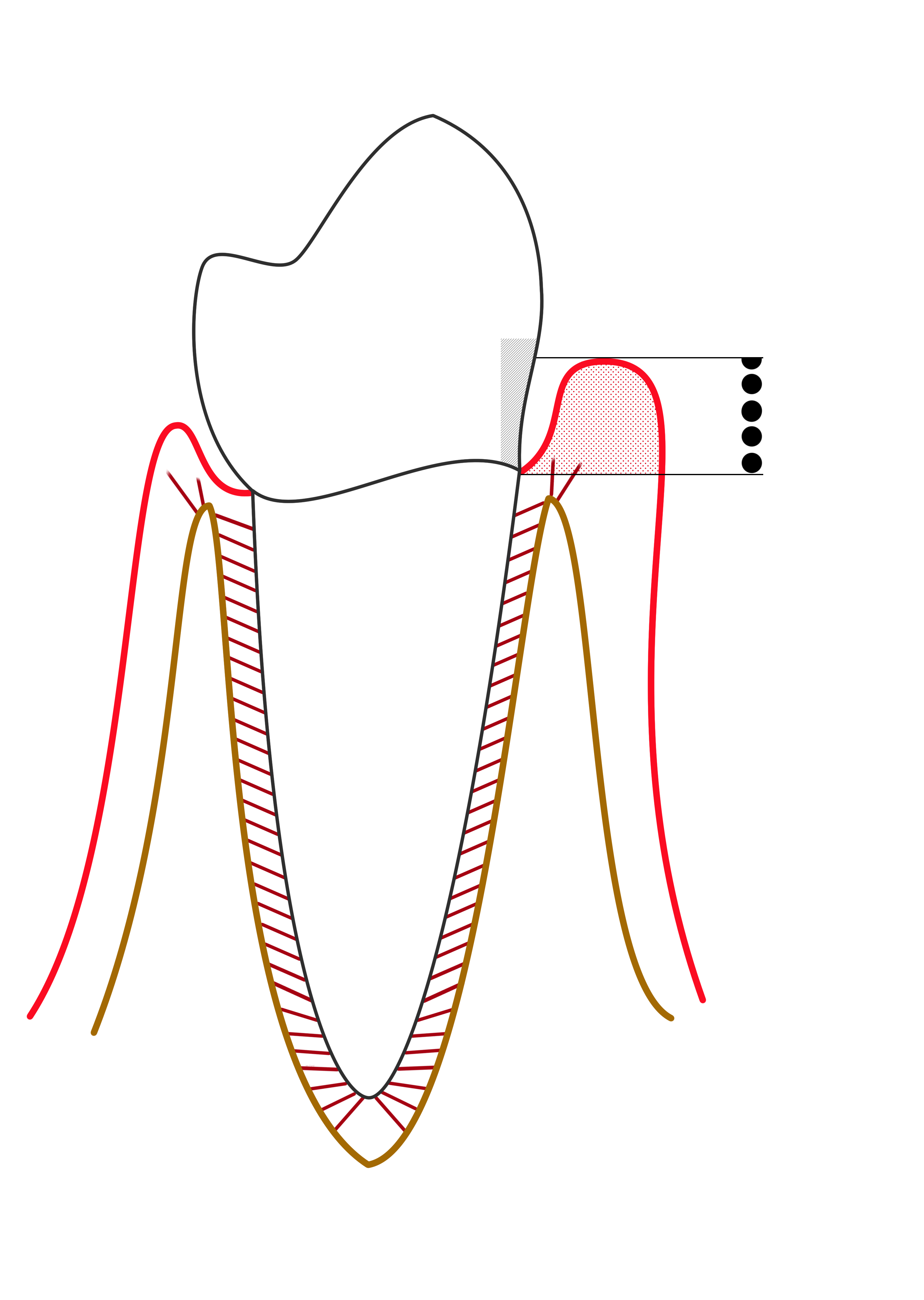
The red marginal outline represents gingival inflammation. Compare the gingival margin, sulcus and supporting bone with the periodontitis diagram. The probe-scale dots illustrate depth, not a diagnosis by themselves.
Line diagram of Gingivitis.png · Ian Furst · CC BY-SA 4.0. None; reproduced without alteration.
Does gingival bleeding alone establish periodontitis?
No. Periodontitis requires evidence of supporting attachment loss, interpreted with clinical and radiographic findings.
Health can exist on a reduced periodontium
An intact periodontium has no previous periodontitis-related attachment or bone loss. A reduced periodontium may follow traumatic recession, surgery or successfully treated periodontitis. These histories are different even when the gingivae look similar today. A treated periodontitis patient retains susceptibility and needs ongoing periodontal monitoring.
Assess bleeding across the mouth rather than judging health from one papilla’s colour. A bleeding score below 10% supports clinical gingival health in the appropriate context. Gingivitis is commonly described as localised at 10–30% bleeding sites and generalised above 30%. These percentages describe inflammation, not the distribution of historical bone loss.
Periodontitis: pocket formation and loss of support
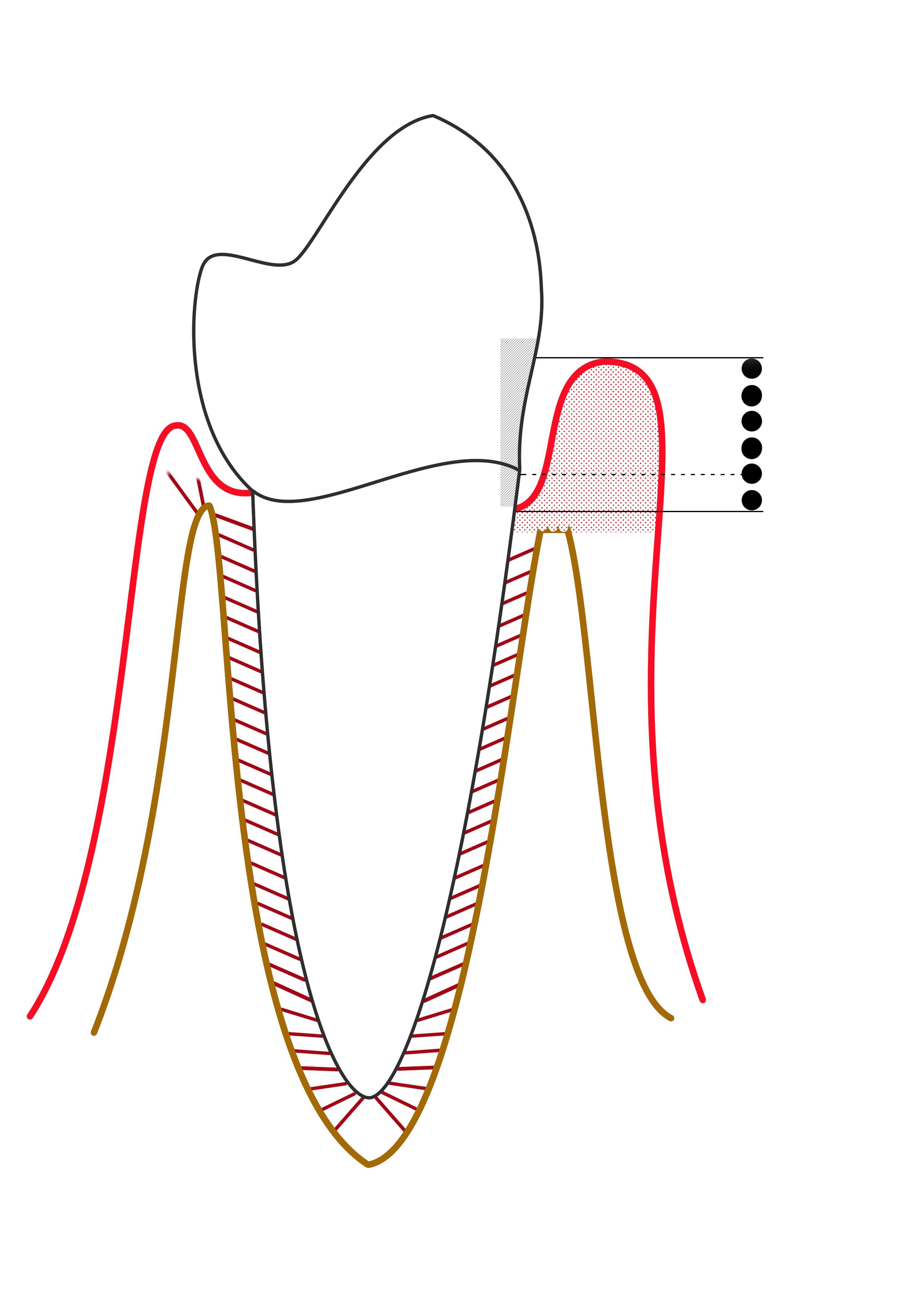
Compare the apical extent of the pocket and the reduced supporting bone on the affected side. This simplified diagram is labelled “mild periodontitis” by its author; it is not a modern stage/grade classification chart.
Mild periodontitis.png · Ian Furst · CC BY-SA 4.0. None; reproduced without alteration.
Why is probing depth different from clinical attachment level?
Probing depth runs from the gingival margin to the pocket base. Clinical attachment level uses the cemento-enamel junction as its reference; recession or enlargement changes their relationship.
Pocket depth is not attachment level
Probing depth runs from the current gingival margin to the pocket base. Clinical attachment level uses a stable reference, usually the cemento-enamel junction. A 3 mm pocket with 4 mm recession can therefore have more attachment loss than a swollen 5 mm gingival pocket without apical attachment loss. Record the margin position rather than interpreting pocket depth in isolation.
Radiographs show mineralised support and historical destruction; they do not measure the gingival soft-tissue pocket. Inflammation around teeth with old bone loss must be interpreted against that history. The phrase healthy gums should not imply that previously destroyed supporting tissues have returned.
Recognise modifiers and alternative disease
Pregnancy and puberty can exaggerate plaque-related inflammation. Some calcium-channel blockers, ciclosporin and phenytoin can be associated with enlargement. Control local inflammation and liaise with the prescriber if a medicine change is considered; do not advise unilateral cessation. Dry mouth and awkward restorations may also make an established cleaning routine less effective.
Persistent desquamation, ulceration, unusual focal enlargement or disproportionate spontaneous bleeding needs a wider differential. Consider mucocutaneous disease, haematological illness and neoplasia when the findings warrant it. Painful necrotic papillae with spontaneous bleeding are different from routine painless gingivitis. Examine the rest of the mucosa and ask about systemic symptoms.
Make plaque control practical
Show the patient the sites being missed and help them practise reaching the gingival margin. Choose interdental aids for the actual spaces and dexterity. Demonstrating a brush size and direction is more useful than simply recommending floss or a branded product. Remove deposits and correct barriers that prevent effective cleaning.
A short-term antiseptic rinse may help selected circumstances, but it neither substitutes for mechanical cleaning nor explains persistent bleeding. Discuss product-specific adverse effects such as staining where relevant. Professional cleaning supports daily care; it cannot permanently compensate for an inaccessible margin or a technique that the patient cannot reproduce.
Review the response and keep the diagnosis accurate
Reassess plaque, bleeding, swelling and symptoms at a suitable interval. Improvement supports a plaque-induced process. Residual pockets require review of attachment, local anatomy and whether periodontitis is present. Record site-level findings where needed so a future clinician can understand what changed.
For example, an adult with crowded lower incisors may improve after calculus removal and tailored interdental instruction. If bleeding settles but interdental recession and bone loss remain, describe controlled inflammation on a reduced periodontium. Shallow non-bleeding pockets are welcome, but they do not erase the disease history or remove the need for supportive care.
A distinction to remember
Bleeding describes current inflammation; it does not establish how much periodontal support has already been lost.
A tooth has a 3 mm pocket and 4 mm recession. Is the attachment level normal?
Reveal the reasoning
No. If the gingival margin is 4 mm apical to the CEJ, the attachment measurement is approximately 7 mm. Establish the cause and distribution of the loss. A shallow pocket alone does not prove that the original periodontal support is intact.
Test your recall
What distinguishes gingivitis from periodontitis?
Show answer
Gingivitis is inflammation confined to the gingiva without periodontitis-related attachment loss. Periodontitis includes destruction of supporting attachment and bone. Assess the history as well as today’s findings, because a treated periodontitis patient can have little inflammation while retaining a reduced periodontium.