
Orthodontic records
Records should answer a planning question and make change measurable. A complete file combines clinical findings with proportionate photographs, models and justified imaging; a scan alone cannot substitute for examination.
Quick revision summary
Records should answer clinical questions
History, examination, photographs and models or scans have complementary roles. Collect sufficient information to diagnose, plan, communicate and review treatment, with secure storage and appropriate consent for each use.
Justify each radiographic exposure
Orthodontic treatment does not justify an automatic set of images. Choose views for the specific question, use available adequate records where appropriate, and report the whole image rather than only tracing teeth.
Use measurements with clinical judgement
Cephalometric values describe selected relationships and depend on landmarks and reference assumptions. Interpret them with facial assessment, growth and the patient’s goals rather than allowing one angle to determine treatment.
Common mistake
A digital treatment animation is a proposed setup. It is not proof of safe root movement, periodontal support or an achievable final bite.
Orthodontic records
Records should answer a planning question and make change measurable. A complete file combines clinical findings with proportionate photographs, models and justified imaging; a scan alone cannot substitute for examination.
Begin with a structured clinical record
Record the patient’s concern, relevant medical and dental history, previous trauma, treatment, habits and family or growth information. Describe facial, skeletal and occlusal findings, oral-health status and the problem list. Distinguish what the patient requests from what treatment can reasonably achieve. Note any time-sensitive eruption or periodontal concern.
The record should explain why the plan was chosen, including alternatives, risks, no treatment and any limits accepted by the patient. Record the consent discussion, rather than relying on a signature or appliance order. At later visits document movement, tissue health, instructions, breakages, changes to the plan and the next review responsibility.
Use standardised photographs for comparison
Extraoral views commonly document frontal appearance at rest and smiling, profile and relevant asymmetry. Intraoral views show the anterior bite, right and left buccal segments and upper and lower arches. Consistent posture, lighting, scale and magnification make baseline and progress images more comparable. Photographs should show the finding without manipulation that changes the clinical impression.
A photograph cannot demonstrate every root, functional contact or periodontal pocket. Supplement it with examination and other records where needed. Obtain appropriate consent, explain purpose and storage and use an approved secure system. Clinical consent does not automatically permit social-media advertising, teaching or publication. A smiling frontal image may identify a person even when the eyes are hidden.
Understand what models and scans contribute
Study models or digital scans can record tooth shape, contact relationships, crowding, arch form, occlusion and space. A digital setup can illustrate proposed movement, but it is a simulation informed by assumptions. It cannot show that a root will safely follow the crown or that the periodontal envelope can tolerate the proposed change.
Check the accuracy of the bite registration and scan completeness, especially distal teeth and gingival detail. An apparently open bite in a scan may reflect acquisition error. Retain versioned baseline and progress records so that the original condition remains available. Appliance manufacture from a model does not transfer diagnostic responsibility to software or a laboratory.
Justify radiographs for a specific need
A panoramic image may help assess the presence, position and development of teeth when this affects planning. Periapicals give more detailed root or periapical information in selected sites. A lateral cephalogram can support assessment of skeletal and incisor relationships when the result will influence treatment. None is mandatory merely because a patient has an orthodontic appointment.
Check existing images for adequacy and obtain additional exposure only where the expected benefit justifies it. Select the smallest appropriate field and technique. CBCT is not routine orthodontic screening: it may be indicated for a specific three-dimensional issue that conventional images cannot resolve adequately. Report the full relevant image, not only the tooth that prompted it.
Lateral cephalogram: orient the orthodontic record
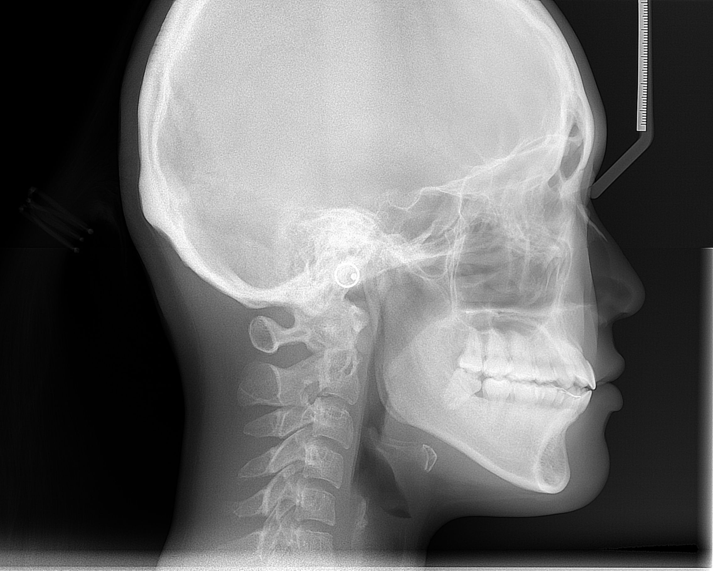
Orient the cranial base, jaws, incisors and soft-tissue outline. Use this as an anatomy example alongside the chapter on justification and interpretation of orthodontic records.
Cephalometric radiograph.JPG · ANUG · CC BY-SA 4.0. Image reproduced without alteration; original marks and any embedded credits retained.
Interpret measurements in clinical context
Cephalometric landmarks and angles summarise relationships, but are affected by landmark identification, projection and individual anatomy. A single ANB value is not the whole skeletal diagnosis. Combine measurements with facial appearance, vertical pattern, dental compensation, growth and the patient’s goals. Norms describe a reference population rather than a compulsory target for every face.
Space calculations and predicted tooth sizes also involve assumptions. Check whether an apparent discrepancy comes from crowding, tooth-size variation, rotations, migration or missing teeth. Repeated records should have a reason, such as suspected resorption or a change in the plan. More measurements do not automatically create more certainty if the underlying record is inaccurate.
Maintain continuity, security and traceability
Identify images and scans with the correct patient and date, record the indication and findings, and ensure they remain accessible to authorised clinicians. Correct errors in a traceable way; do not silently overwrite the original account. Transfer records securely with relevant consent and governance, checking the recipient rather than using informal personal messaging.
Retention and disposal must follow the applicable legal, professional and organisational schedule, which can vary with jurisdiction, patient age and circumstances. A device change or software subscription should not make the clinical baseline inaccessible. At transfer, communicate treatment stage, appliances, outstanding concerns, risks and retention plans. A set of photographs without that narrative is an incomplete handover.
A distinction to remember
A digital treatment animation is a proposed setup. It is not proof of safe root movement, periodontal support or an achievable final bite.
A referral arrives with an attractive digital setup but no history or periodontal findings. What is missing before treatment?
Reveal the reasoning
A clinical assessment, diagnosis, baseline tissue and occlusal findings, relevant trauma and medical history, and justified records are missing. Confirm the setup’s assumptions and the patient’s goals and consent before selecting mechanics. Software output cannot establish biological suitability.
Test your recall
What should an orthodontic baseline record explain?
Show answer
It should explain the patient’s concerns, diagnosis, relevant risk factors, clinical findings and the reasons for the chosen plan. Include alternatives, consent and goals. Images and measurements support this account, but do not replace the decision-making narrative.