
Obturation and coronal seal
Canal filling and definitive restoration are linked stages of infection control and tooth preservation.
Quick revision summary
Assess the canal treatment and the coronal restoration
Obturation aims to seal the prepared canal system after appropriate treatment. Interpret symptoms, canal condition, anatomy and procedural findings before filling.
Decide whether the canal is ready to fill
Completion depends on the diagnosis, treatment performed and current canal conditions. Persistent drainage, unexplained symptoms or uncertainty about anatomy should be assessed before simply proceeding to obturation.
Protect the tooth and judge healing over time
Plan the definitive coronal restoration early enough to avoid prolonged reliance on a leaking or fragile temporary seal. Remaining walls, marginal ridges, cracks, occlusal loading and the proposed restoration influence the need for cuspal protection.
Common mistake
A good-looking root filling cannot rescue a tooth that is leaking, fractured or unrestorable.
Obturation, coronal seal and outcome
Canal filling and definitive restoration are linked stages of infection control and tooth preservation.
Assess the canal treatment and the coronal restoration
Obturation aims to seal the prepared canal system after appropriate treatment. Interpret symptoms, canal condition, anatomy and procedural findings before filling. A dense radiographic appearance is only a projected technical observation; it cannot prove the whole system is disinfected or every recess is sealed. Sealer is not a substitute for sound preparation and a suitable filling technique.
The coronal restoration prevents reinfection and restores the tooth’s structural function. Plan its timing and design from remaining tissue, occlusion and prognosis. A temporary restoration has a purpose and an endpoint; prolonged leakage can undermine previous treatment. Follow-up combines symptoms, function, examination and radiographic change over time. Healing may take time, so compare with baseline rather than calling every residual shadow immediate failure.
Key distinctions in practice
| Finding or option | What it means | Clinical consequence |
|---|---|---|
| Obturation quality | Technical canal fill | Assess in context of anatomy and infection control. |
| Coronal leakage | Route for reinfection | Provide a reliable restoration and completion plan. |
| Cuspal protection | May reduce fracture risk | Choose from remaining tissue and load. |
| Serial radiography | Shows the healing trajectory | Interpret with clinical response and elapsed time. |
Decide whether the canal is ready to fill
Completion depends on the diagnosis, treatment performed and current canal conditions. Persistent drainage, unexplained symptoms or uncertainty about anatomy should be assessed before simply proceeding to obturation. A filling material cannot compensate for an untreated canal, a perforation or inadequate disinfection. Record procedural findings that affect the interpretation of the finished image.
Assess the resulting fill for its extent, density and relationship to the intended preparation while recognising the limitations of a two-dimensional projection. Extruded material is not evidence of thorough cleaning. A technically satisfactory appearance is one part of the assessment, alongside the biological treatment and the tooth’s capacity to function after restoration.
Root-filled molar: trace the visible canals
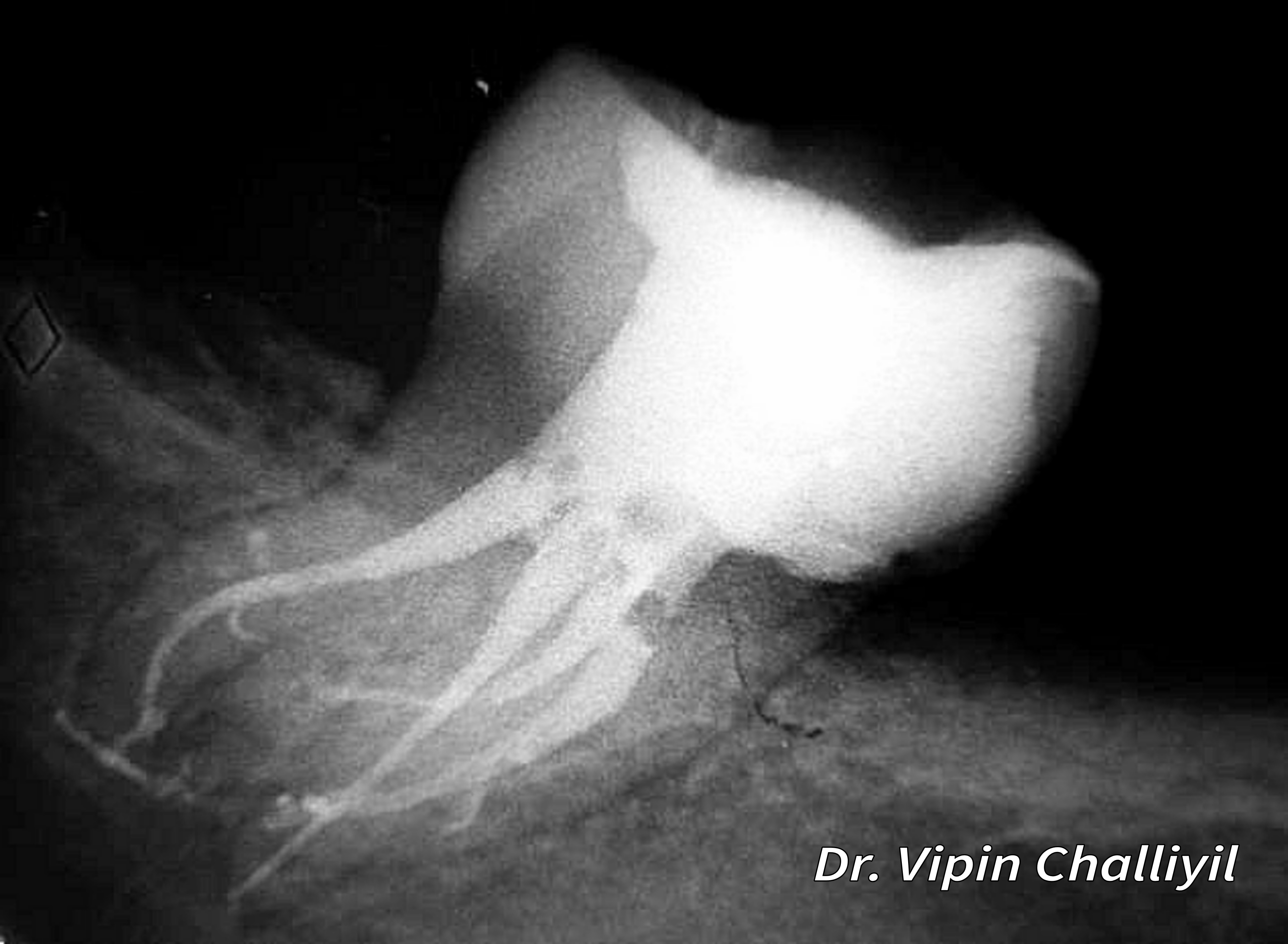
Follow the radiopaque root fillings and the surrounding root outlines. The image illustrates canal anatomy after treatment; radiographic appearance alone does not establish disinfection or healing.
Root canal treatment of lone standing molar.jpg · Challiyan (Dr Vipin Challiyil) · CC BY-SA 4.0. Image reproduced without alteration; original marks and any embedded credits retained.
Protect the tooth and judge healing over time
Plan the definitive coronal restoration early enough to avoid prolonged reliance on a leaking or fragile temporary seal. Remaining walls, marginal ridges, cracks, occlusal loading and the proposed restoration influence the need for cuspal protection. A post may retain a core when needed; placing one does not automatically strengthen a root-filled tooth.
Document baseline symptoms, examination and appropriate postoperative imaging. At review, assess comfort, function, soft tissues and changes in apical findings. A lesion that is decreasing over time differs from one that is enlarging with new symptoms. Persistent radiographic change cannot be labelled solely by how dense the root filling looks. Investigate leakage, missed anatomy, fracture and other sources when the clinical course is unsatisfactory.
A distinction to remember
A good-looking root filling cannot rescue a tooth that is leaking, fractured or unrestorable.
Why is a good-looking root filling insufficient if the crown restoration leaks?
Reveal the reasoning
Coronal contamination can reintroduce microbes and the tooth may remain mechanically vulnerable. Endodontic and restorative quality work together.
Guidance to read alongside this chapter
Test your recall
Is obturation the only determinant of success?
Show answer
No; disinfection, anatomy and the definitive coronal seal also matter.