
Spreading infection
Assess the airway, the rate and anatomical route of spread, and the patient’s physiology. Antibiotics, drainage and emergency support solve different problems; a prescription is not a substitute for identifying which of them is needed.
Quick revision summary
Assess beyond the tooth
Examine breathing, voice, swallowing, mouth opening, floor of mouth, neck and systemic state. Deep-space infection may be more extensive than the visible swelling, particularly in an immunocompromised person or with limited examination.
Escalate dangerous spread
Airway threat, rapidly progressive swelling, severe trismus, systemic deterioration or eye involvement requires urgent appropriate hospital assessment. Do not delay escalation for routine radiography or a trial of another oral antibiotic.
Combine source control and review
Drainage or treatment of the causal tooth is central when feasible. Antibiotics have defined indications in spreading or systemic infection; document the diagnosis, host factors, intervention and reassessment rather than treating the prescription as definitive care.
Common mistake
A normal temperature or low early-warning score does not exclude a threatened airway or serious infection in an immunocompromised patient.
Spreading infection
Assess the airway, the rate and anatomical route of spread, and the patient’s physiology. Antibiotics, drainage and emergency support solve different problems; a prescription is not a substitute for identifying which of them is needed.
Understand the route beyond the tooth
Pulpal or periodontal infection can breach bone and involve adjacent soft tissues. The relation of the root apex and cortical perforation to muscle attachments influences the initial space involved. Lingual spread above the mylohyoid may enter the sublingual space; spread below can involve the submandibular region. Buccinator attachment influences vestibular versus buccal involvement. Masticator-space disease can cause substantial trismus. These relationships help explain the pattern but should not delay assessment of airway and circulation. A small visible intraoral swelling may underestimate deeper disease, particularly when access is limited or the host response is impaired.
Mandibular landmarks
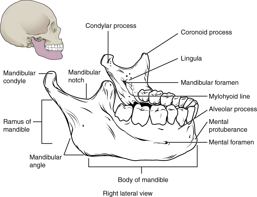
Locate the mental and mandibular foramina, lingula and mylohyoid line. The mandibular foramen, lingula and mylohyoid line are medial landmarks; this schematic brings several surfaces together. Relate the landmarks to nerve risk and fracture assessment.
Mandibular landmarks · OpenStax College · CC BY 3.0. Original image; displayed at a responsive size. Original publication.
Examine the patient, not just the swelling
Ask about onset, progression, dental source, previous drainage, antibiotics and host factors. Assess breathing, speech, voice, swallowing, drooling, mouth opening, tongue position, floor of mouth, neck and eye symptoms where relevant. Record observations and mental state when systemic illness is possible. Palpate and inspect carefully without forcing a patient with airway concern into an intolerable position. Determine whether swelling is localised and fluctuant or diffuse and spreading, but do not assume absence of fluctuation means absence of a serious infection. Examine possible source teeth with appropriate imaging only when the patient’s stability allows it.
Coronal sublingual and submandibular spaces with normal MRI correlation
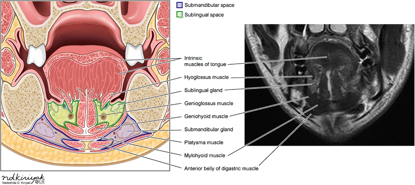
Identify the mylohyoid and compare the sublingual and submandibular compartments in the schematic and normal MRI. Relate the compartments to the tooth-apex relationships discussed in this section.
Coronal sublingual and submandibular spaces with normal MRI correlation · Swapnil Patel and Alok A. Bhatt (2018); illustration credited within the figure to Nadezhda D. Kiriyak / University of Rochester · CC BY 4.0. Unchanged published image; displayed responsively. Original publication.
Recognise threatened airway and deep spread
Difficulty breathing, inability to manage secretions, altered voice, tongue elevation and bilateral floor-of-mouth or neck swelling are concerning. Significant trismus, rapidly progressive cellulitis or deep-space features can indicate disease unsuitable for repeated oral prescriptions. Call emergency ambulance support for threatened airway or sepsis in community dental care; activate the local emergency response in hospital. Arrange urgent surgical and airway expertise and communicate the pattern clearly. Do not delay transfer while seeking a perfect fascial-space label, performing a difficult radiograph or waiting for antibiotics to work. Airway management must fit the anatomy and expertise available.
Suprahyoid and infrahyoid muscles
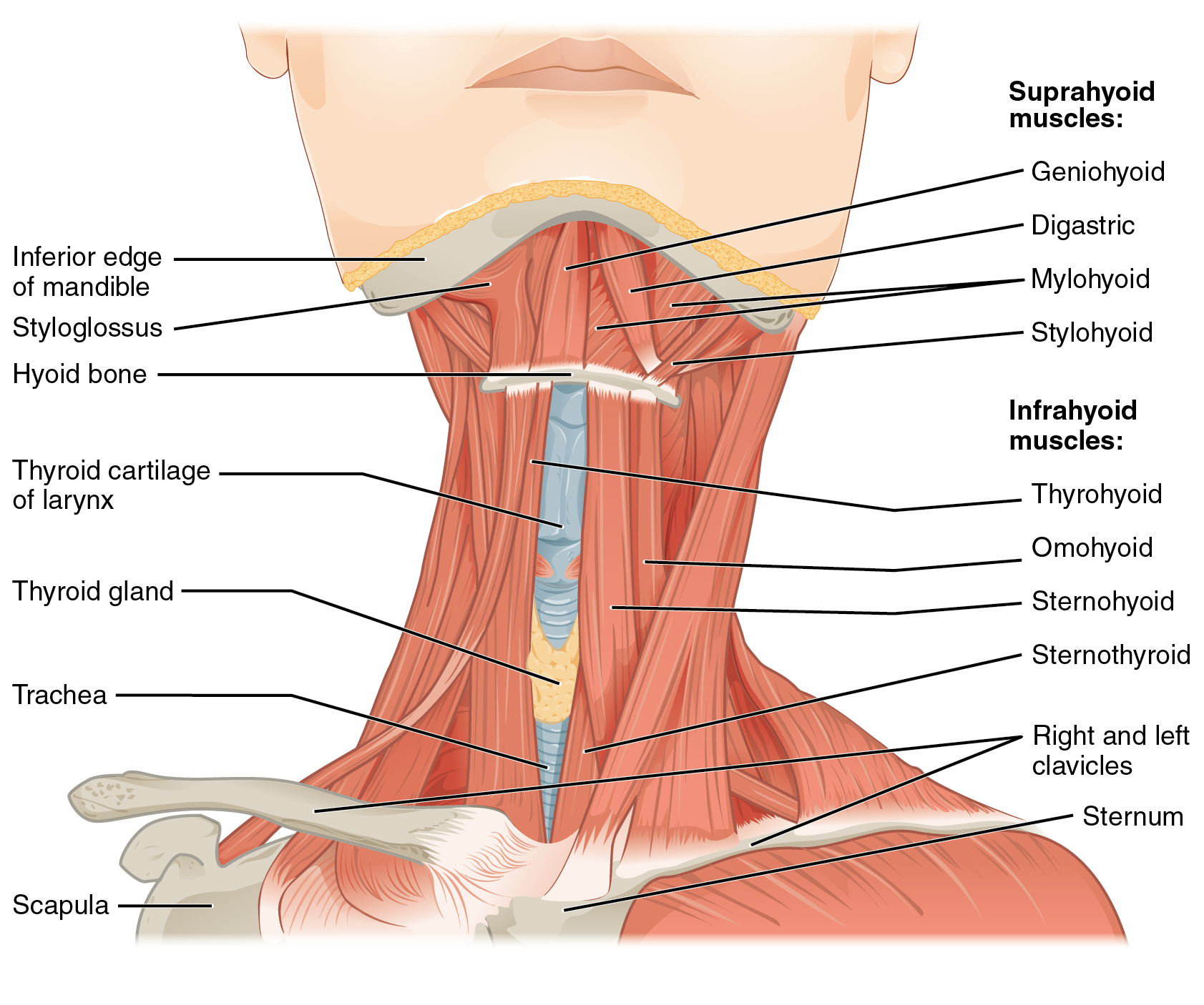
Locate the hyoid, then the four suprahyoid muscles. The mylohyoid forms the muscular floor of the mouth; the geniohyoid lies above it. Relate these muscles to swallowing, mandibular depression and the sublingual–submandibular boundary. The figure shows selected layers exposed for teaching.
1110 Muscle of the Anterior Neck.jpg · OpenStax · CC BY 4.0. None; original image reproduced without alteration.
Which muscle forms the main muscular floor of the mouth?
Mylohyoid. Geniohyoid lies superior to it; the anterior belly of digastric lies inferior to it.
Assess possible sepsis using the right pathway
Sepsis involves life-threatening organ dysfunction associated with infection and can occur without high fever. New confusion, abnormal respiration, poor perfusion and a marked change from usual function matter. NICE NG253 applies to people aged 16 or over who are not and have not recently been pregnant; use the relevant separate pathway for children or pregnancy. NEWS2 supports assessment in its specified acute settings, while community assessment has its own recommendations. A score cannot override an anatomical airway threat or clinical concern. Record and repeat observations where appropriate because deterioration may occur while arrangements are being made.
Obtain source control and use antibiotics for an indication
A localised abscess generally needs drainage and removal of the cause through appropriate dental treatment. Spreading infection, systemic involvement or inability to achieve immediate drainage may require antibiotics alongside a definite source-treatment plan. The route, agent and setting depend on severity, allergies, interactions and local guidance. Severe disease may need hospital admission, intravenous therapy and operative drainage. Avoid blind incision into an undiagnosed diffuse swelling or an unsafe space. Culture or imaging can support management when indicated, but neither should postpone emergency intervention in an unstable patient or replace elimination of the source.
Investigate according to stability and the clinical question
Dental imaging helps identify the source and anatomy; cross-sectional imaging can help define deep spread or a collection in a stable patient under the appropriate team. Blood tests, cultures and lactate can contribute to systemic assessment, but no isolated result determines airway safety. Consider immune status, renal function and previous treatment when interpreting the picture and planning medicines. A patient on anticancer therapy who becomes unwell may need the neutropenic-sepsis pathway even if the mouth looks unimpressive. A normal temperature or a modest inflammatory marker must not become a reason to ignore an obviously deteriorating patient.
Review response and give an executable escalation plan
Explain what improvement is expected, how the source will be treated and where to seek urgent help. SDCEP recommends antibiotic review ideally at three days, with earlier reassessment for rapid worsening, new symptoms or failure to improve. At review, reconsider the diagnosis, drainage, medicine use, host factors and anatomical spread before changing antibiotics. When systemic signs have resolved, follow current advice on stopping rather than automatically prolonging treatment. Communicate with the receiving service using the suspected source, progression, airway findings, observations, medicines and actions already taken. Ensure someone owns the next step rather than merely issuing an untracked referral.
A distinction to remember
A normal temperature or low early-warning score does not exclude a threatened airway or serious infection in an immunocompromised patient.
A patient has lower-molar pain, a raised floor of mouth, drooling and a muffled voice. Is another oral antibiotic course appropriate?
Reveal the reasoning
This is an emergency pattern suggesting deep spread and possible airway compromise. Arrange emergency transfer and specialist airway and surgical support, with ABCDE care while waiting. Do not delay for routine imaging or expect an oral prescription to secure the airway.
Test your recall
Which anatomical relationship helps explain sublingual versus submandibular spread?
Show answer
The point of lingual cortical perforation relative to the mylohyoid attachment influences the initial space involved. This helps interpret swelling, but spaces communicate and progression varies. Anatomy supports clinical assessment and does not take priority over an airway threat.