
Core practical skills
Bloods, cannulation, suturing and local anaesthesia within competence.
Clinical images and diagrams
Trigeminal divisions and facial sensory territories
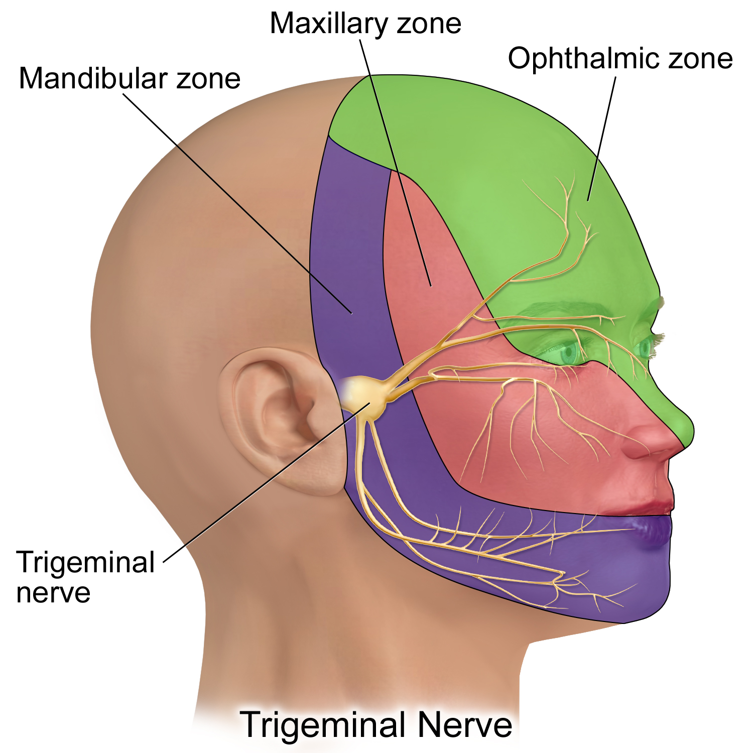
V1 is ophthalmic, V2 maxillary and V3 mandibular. This is a schematic sensory territory map, not a detailed map of every peripheral branch; V3 also carries motor fibres.
Trigeminal Nerve.png · BruceBlaus · CC BY-SA 4.0. None; reproduced without alteration.
Which division gives rise to the inferior alveolar and lingual nerves?
The mandibular division, V3. Distinguish lower lip/chin sensory change from altered tongue sensation.
What to expect
Skills often include venepuncture, peripheral cannulation, wound assessment, local anaesthesia and selected suturing under supervision. Opportunities vary. Build competence through observed practice and local training rather than treating a presentation as authorisation to perform a procedure.
What to know
For bloods, match the request to the clinical question and use the correct identification, collection and labelling process. For a cannula, consider indication, site, infection prevention, patency, documentation and daily need. A VBG may help assess acid-base status, lactate or electrolytes in an unwell patient when ordered under the local pathway; an ABG is usually a more specialist decision for oxygenation or severe respiratory assessment. Understand the limits of each sample. For wounds, assess depth, contamination, tissue viability, nerves, ducts and vessels before closure.
A practical approach
Explain the procedure, check consent and allergies, prepare equipment and sharps safety, then work with supervision. Record attempts, site, sample or device details, complications and aftercare. For sutures, aim for accurate alignment without undue tension and arrange review/removal under local protocol.
Safety point
Do not close a facial wound before assessing deeper injury or persist with repeated unsuccessful access. Escalate when anatomy, patient condition or complexity exceeds your training.
Peripheral venous access and cannulation
Know the anatomy, practical checks and complications of vascular access while working within your training and local procedure.
Upper-limb vascular access
The cephalic vein lies laterally, the basilic vein medially and the median cubital vein commonly connects them in the antecubital region. Patterns vary. A palpable pulse, unusually bright pulsatile return, marked pain, swelling or resistance is not a finding to ignore. Nearby arteries and cutaneous nerves make anatomical awareness and careful site selection necessary.
Choose a suitable superficial vein, use aseptic technique, confirm satisfactory placement and secure the cannula without hiding the site. Pain or swelling during administration suggests extravasation or another placement problem: stop administration and assess. A cannula that originally worked can subsequently dislodge. In sedation, monitoring must include the patient and the cannulation site, not just a monitor display.
Superficial and deep veins of the upper limb
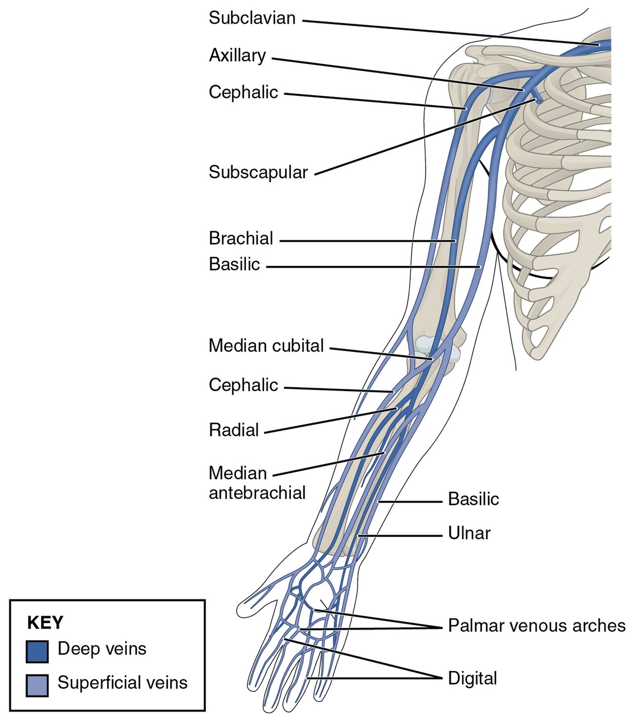
Identify cephalic, basilic, median cubital and median antebrachial veins. Use the colour key to distinguish superficial and deep drainage, and relate the anatomy to venous-access assessment. Individual superficial-vein patterns vary.
Superficial and deep veins of the upper limb · OpenStax College (2013) · CC BY 3.0. Unchanged published raster preview; displayed responsively. Original available from the source. Original publication.
Confirm function and watch the site
Use the local aseptic technique and equipment procedure, suitable patient positioning and appropriate supervision. Identify a suitable vein and consider contraindicated or compromised sites according to the patient and local policy. Explain the procedure and record the access, attempts and any complication. A diagram supports anatomical orientation but cannot establish procedural competence.
Check and secure the access using the appropriate local protocol and reassess during use. Pain, swelling, resistance, leakage or altered function can indicate a problem. Stop administration and assess rather than forcing an injection or relying on an earlier blood return. Manage suspected extravasation through the medicine-specific and local pathway and obtain senior help when needed.
Trigeminal divisions and facial sensory territories
V1 is ophthalmic, V2 maxillary and V3 mandibular. This is a schematic sensory territory map, not a detailed map of every peripheral branch; V3 also carries motor fibres.
Trigeminal Nerve.png · BruceBlaus · CC BY-SA 4.0. None; reproduced without alteration.
Which division gives rise to the inferior alveolar and lingual nerves?
The mandibular division, V3. Distinguish lower lip/chin sensory change from altered tongue sensation.
Access requires continued assessment
Before
- Confirm indication, consent and suitable site
- Use trained aseptic technique
During use
- Confirm and secure access
- Observe site and patient during administration
If function changes
- Stop administration and reassess
- Follow the complication pathway and seek help
Use the trust’s practical procedure and medicine-specific advice; anatomical knowledge alone does not confer competence.
A distinction to remember
A previous blood return does not guarantee that subsequent administration remains intravascular.
A sedated patient develops swelling at the cannula while a medicine is injected. What is the immediate change?
Reveal the reasoning
Stop administration, assess the site and patient, and follow the extravasation / cannulation pathway. Do not inject further merely because the cannula gave a satisfactory blood return earlier.
Guidance to read alongside this chapter
Biopsy technique, sutures and operative equipment
Good tissue handling protects both healing and diagnostic value. Understand what each tool does and the additional hazards it introduces.
Biopsy is a planned diagnostic operation
An incisional biopsy samples a representative part of a larger or suspicious lesion; an excisional biopsy removes an appropriately selected lesion. The choice depends on size, suspected pathology, anatomical risk and definitive treatment planning. Avoid removing a suspected malignancy casually without considering orientation, margins and the cancer pathway.
Take viable tissue with adequate depth and minimise crush or thermal artefact. Arrange separate specimen handling when immunofluorescence or microbiology is required. Label accurately and include the clinical history, site, medicines and differential. A technically easy sample can still be diagnostically poor if it is taken from the wrong area.
Inflammation, granulation and maturation in tissue repair
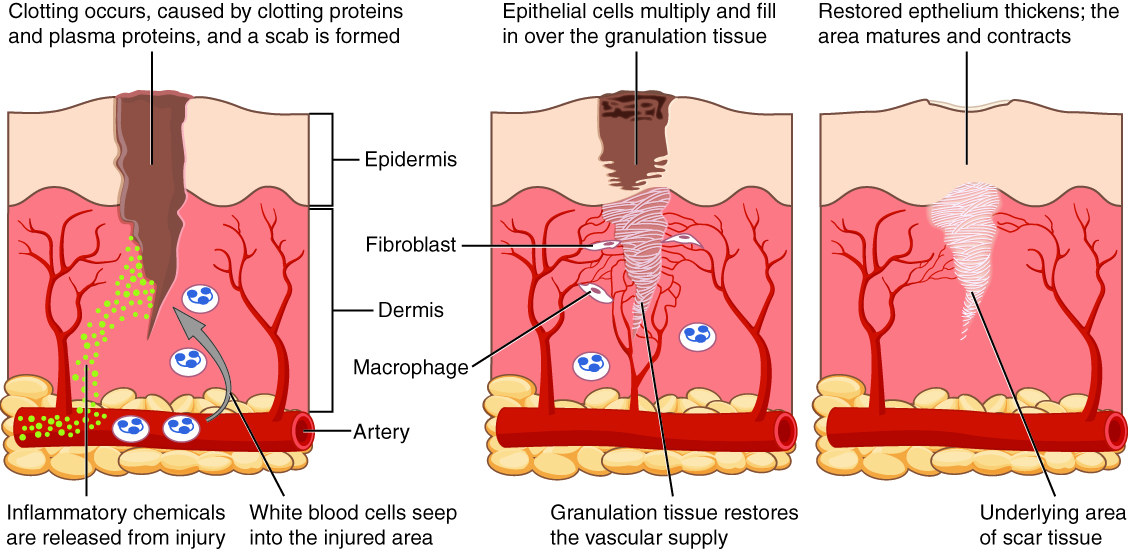
Relate inflammatory cells, fibroblasts, granulation tissue, epithelialisation and scar maturation to the overlapping stages of wound healing. This is a skin-wound example, not an extraction socket; oral mucosa differs and does not necessarily form a dry scab. Healing phases overlap.
Inflammation, granulation and maturation in tissue repair · OpenStax College (2013) · CC BY 3.0. Unchanged published image; displayed responsively. Original publication.
Equipment and material choices
| Tool / material | Mechanism / benefit | Specific consideration |
|---|---|---|
| Monofilament suture | Smooth single strand; limited wicking | Handling and knot security differ from braided material. |
| Braided suture | Useful handling and knot characteristics | Tissue response and wicking / plaque retention matter. |
| Absorbable suture | Loses strength / breaks down over time | Strength retention must fit the tissue and healing requirement. |
| Diathermy | Electrical energy used for cutting / coagulation | Thermal spread, device interactions and appropriate circuit / safety precautions. |
| Laser | Wavelength-dependent interaction with tissue | Eye protection, smoke, thermal injury and specimen artefact. |
| Piezoelectric bone surgery | Ultrasonic cutting of mineralised tissue | Irrigation, tip use, heat and operative time still require control. |
Trigeminal divisions and facial sensory territories
V1 is ophthalmic, V2 maxillary and V3 mandibular. This is a schematic sensory territory map, not a detailed map of every peripheral branch; V3 also carries motor fibres.
Trigeminal Nerve.png · BruceBlaus · CC BY-SA 4.0. None; reproduced without alteration.
Which division gives rise to the inferior alveolar and lingual nerves?
The mandibular division, V3. Distinguish lower lip/chin sensory change from altered tongue sensation.
Principles that apply to every technique
Maintain visibility, protect adjacent structures and match energy or mechanical force to the task. More technologically advanced equipment does not remove the need for anatomy or technique. Equipment training, maintenance and the local safety procedure are part of choosing the device.
Select sutures for the tissue, tension, contamination, strength requirement and follow-up. Approximate rather than strangulate tissue; excessive tension and multiple traumatic passes impair healing. Document specimen disposition, any equipment issue and the wound plan, and ensure the histology result is reviewed and communicated.
Choose the tool for the biological task
Diagnostic tissue
- Select representative site and depth
- Minimise crush and thermal damage
- Use correct laboratory handling
Wound approximation
- Assess tissue and tension
- Choose suitable material / technique
- Preserve perfusion during healing
Energy device
- Know mechanism and hazards
- Protect eyes, tissue and equipment safety
- Use training and local procedure
A distinction to remember
A laser or diathermy margin can create artefact in a diagnostic specimen. Haemostasis should not destroy the tissue needed to answer the pathological question.
Why is a necrotic ulcer base alone often a poor biopsy sample?
Reveal the reasoning
It may contain debris and inflammation without the viable lesional architecture needed for diagnosis. Select a representative viable area and adequate depth, with handling appropriate to the suspected condition.
Test your recall
What is the key limitation of a VBG compared with an ABG?
Show answer
A VBG does not directly assess arterial oxygenation; the test must match the clinical question.